
Best Orthopedic Surgeon for ACL Surgery in Hyderabad

Knee injuries are among the most common orthopedic problems, especially in athletes, active individuals, and people involved in physically demanding activities. One of the most serious knee injuries is an Anterior Cruciate Ligament (ACL) tear, which can lead to pain, instability, and difficulty performing everyday activities. If you are searching for the Best Orthopedic Surgeon for ACL Surgery in Hyderabad, consulting an experienced specialist is essential for proper diagnosis and successful treatment. Dr. Arun Reddy Mallu is a highly skilled orthopedic and sports injury specialist associated with Continental Hospitals. He is known for providing advanced treatment for ACL injuries and other knee-related conditions. With modern surgical techniques and personalized patient care, Dr. Arun Reddy Mallu at Continental Hospitals in Hyderabad helps patients regain knee strength, mobility, and confidence after ACL injuries. Understanding ACL Injury The Anterior Cruciate Ligament (ACL) is one of the key ligaments in the knee joint. It connects the thigh bone (femur) to the shin bone (tibia) and helps stabilize the knee during movements such as running, jumping, and sudden direction changes. When this ligament is torn or severely damaged, the knee may become unstable and painful. ACL injuries are very common among athletes, but they can also occur due to accidents or sudden twisting movements. ACL injuries may be classified as: Grade 1:Mild stretching of the ligament Grade 2:Partial tear of the ligament Grade 3:Complete tear of the ligament Severe ACL injuries usually require surgical reconstruction to restore knee stability. Common Causes of ACL Tears Several factors can lead to ACL injuries. Some of the most common causes include: Sudden changes in direction while running Incorrect landing from jumps Sports injuriesduring football, basketball, or cricket Direct impact or collision during sports Road accidents or traumatic injuries Weak muscles around the knee joint Athletes and active individuals are more prone to ACL injuries because their knees experience frequent stress and movement. Symptoms of ACL Injury Recognizing ACL injury symptoms early can help prevent further knee damage. Some common signs include: A sudden popping soundin the knee at the time of injury Severe knee pain immediately after the injury Swelling around the knee joint within a few hours Difficulty walking or putting weight on the leg Knee instability or the feeling that the knee may give way Reduced range of motion If you experience these symptoms, it is important to consult an experienced orthopedic specialist Patients looking for the Best Orthopedic Surgeon in Hyderabad can consult Dr. Arun Reddy Mallu at Continental Hospitals, for proper evaluation and advanced ACL treatment. Diagnosis of ACL Injuries Proper diagnosis is crucial to determine the severity of the injury and the most effective treatment plan. An orthopedic surgeon may perform the following: Physical Examination The doctor checks knee stability, swelling, and range of motion. Imaging Tests Advanced imaging techniques help confirm the diagnosis: MRI Scan:Provides detailed images of ligaments and soft tissues X-rays:Helps rule out bone fractures Arthroscopic evaluation:Sometimes used to examine internal knee structures After evaluating the results, the orthopedic surgeon decides whether non-surgical treatment or ACL reconstruction surgery is required. What Is ACL Reconstruction Surgery? ACL reconstruction surgery is a procedure performed to replace the torn ligament with a tendon graft. The graft functions as a new ligament that restores stability to the knee joint. Modern ACL surgeries are usually performed using arthroscopic techniques, which involve small incisions and specialized surgical instruments. This minimally invasive approach allows faster healing and reduced post-operative discomfort. ACL surgery is recommended for: Complete ACL tears Chronic knee instability Athletes who want to return to sports activities Patients with combined ligament injuries Types of ACL Reconstruction Techniques Different graft options may be used during ACL surgery depending on the patient’s condition. Hamstring Tendon Graft A small portion of the hamstring tendon is used to reconstruct the ACL. This is one of the most common methods. Patellar Tendon Graft A section of the tendon connecting the kneecap to the shin bone is used for reconstruction. Quadriceps Tendon Graft Part of the quadriceps tendon above the kneecap is used to replace the torn ligament. Donor Tissue (Allograft) In some cases, donor tissue may be used instead of the patient’s own tendon. The choice of graft depends on factors such as age, activity level, and the severity of the injury. Why Choose Dr. Arun Reddy Mallu for ACL Surgery in Hyderabad? Choosing the right orthopedic surgeon is crucial for successful ACL treatment and long-term knee health. Dr. Arun Reddy Mallu at Continental Hospitals, Hyderabad is known for his expertise in treating sports injuries and performing advanced knee surgeries. Key Advantages of Consulting Dr. Arun Reddy Mallu Extensive Experience in Orthopedic SurgeryHe has vast experience in diagnosing and treating knee ligament injuries including ACL tears. Advanced Arthroscopic TechniquesModern minimally invasive surgical methods help ensure precise treatment with faster recovery. Patient-Centered CareEvery treatment plan is tailored to the patient’s condition, activity level, and recovery goals. Comprehensive Sports Injury ManagementFrom diagnosis to rehabilitation, patients receive complete care under expert supervision. High Success Rate in ACL ReconstructionProper surgical technique combined with rehabilitation ensures long-term knee stability. Because of these reasons, many patients consider Dr. Arun Reddy Mallu one of the best orthopedic surgeons for ACL surgery in Hyderabad. Recovery and Rehabilitation After ACL Surgery Recovery after ACL surgery is a gradual process that requires proper physiotherapy and rehabilitation. Initial Recovery Phase (First 2 Weeks) Pain and swelling management Use of knee brace or crutches Gentle knee movement exercises Intermediate Phase (2–6 Weeks) Physiotherapy sessions begin Gradual improvement in knee mobility Muscle strengthening exercises Strengthening Phase (6 Weeks to 3 Months) Increased strength training Improved balance and flexibility Advanced Recovery (4–6 Months) Light sports activities may resume Full Recovery (6–9 Months) Return to full sports participation Following the rehabilitation plan recommended by the orthopedic surgeon is essential for successful recovery. Benefits of ACL Reconstruction Surgery ACL surgery offers several long-term benefits, including: Restores knee stability Improves mobility and flexibility Reduces knee pain and swelling Allows patients to return to sports activities Prevents further knee damage Tips to Prevent ACL Injuries
Tech Neck & Office Posture: A Growing Health Concern

In the modern digital era, technology has transformed the way we work, communicate, and live. From computers and laptops to smartphones and tablets, screens have become a central part of everyday life. While these devices have increased productivity and connectivity, they have also introduced new health challenges. One of the most common and rapidly growing problems is Tech Neck, a condition linked to prolonged device use and poor posture. Tech neck and poor office posture are becoming increasingly common among professionals, students, and even teenagers. Many people spend several hours each day sitting in front of screens, often without realizing the strain this puts on their neck, shoulders, and spine. Over time, this can lead to chronic pain, reduced mobility, and long-term musculoskeletal problems. Understanding the causes, symptoms, and prevention strategies for tech neck is essential for maintaining overall health in today’s technology-driven world. What is Tech Neck? Tech neck refers to neck pain and discomfort caused by repeatedly looking down at digital devices for long periods. This posture places significant pressure on the cervical spine, the upper part of the spine that supports the head. In a neutral position, the human head weighs about 10–12 pounds. However, when the head tilts forward to look at a phone or laptop, the pressure on the neck increases dramatically. At steep angles, the effective weight placed on the neck can increase several times, forcing muscles and ligaments to work much harder than they should. Over time, this strain can lead to muscle fatigue, spinal misalignment, and chronic neck pain. Why Tech Neck is Becoming More Common Several lifestyle and workplace changes have contributed to the increasing prevalence of tech neck. Increased Screen Time One of the biggest reasons for tech neck is the dramatic rise in screen time. Many people spend most of their day using computers for work, followed by additional hours on smartphones for social media, entertainment, and communication. Office employees may spend 8 to 10 hours a day in front of computer screens. After work, many continue using digital devices at home, leaving little time for the body to recover. Poor Workplace Ergonomics Many workplaces are not designed with proper ergonomics in mind. Employees often work with laptops placed too low on desks or without proper chair support. These setups force workers to bend their necks forward or slouch while typing. Common ergonomic issues include: Monitors positioned below eye level Chairs without proper lumbar support Desks that are too high or too low Poor keyboard and mouse placement These factors contribute to poor posture and increased strain on the neck and upper back. Sedentary Work Culture Modern jobs often require prolonged sitting with minimal movement. Sitting for extended periods causes muscles to stiffen and weakens the core and back muscles that support proper posture. When combined with poor sitting posture, this sedentary lifestyle increases the likelihood of developing neck and back pain. Excessive Smartphone Use Smartphones are another major contributor to tech neck. Many people frequently look down at their phones while texting, browsing, or scrolling through social media. This constant downward gaze places continuous pressure on the neck. Young adults and teenagers are particularly at risk because they often spend long periods using mobile devices. Common Symptoms of Tech Neck Tech neck symptoms usually develop gradually and may worsen if left untreated. Many individuals initially dismiss these symptoms as temporary discomfort. Common symptoms include: Persistent neck pain or stiffness Shoulder tightness Upper back discomfort Headaches, especially tension headaches Reduced range of motion in the neck Muscle fatigue in the neck and shoulders Tingling or numbness in the arms or hands In some cases, people may also experience eye strain or fatigue due to prolonged screen exposure. If these symptoms continue for extended periods, they can interfere with daily activities and work performance. Long-Term Health Risks of Poor Posture Ignoring tech neck and poor office posture can lead to serious long-term health issues. Spinal Misalignment Forward head posture can gradually change the natural curvature of the spine. This can lead to structural issues such as rounded shoulders or an exaggerated curve in the upper back. Herniated or Compressed Discs Excess pressure on the cervical spine may cause spinal discs to compress or bulge, potentially irritating nearby nerves. This can lead to radiating pain in the shoulders and arms. Chronic Pain Conditions Untreated posture-related strain may develop into chronic neck or back pain, requiring medical treatment and physical therapy. Reduced Mobility Long-term muscle stiffness can reduce flexibility in the neck and upper back, making everyday movements uncomfortable. Nerve Compression When muscles and joints around the cervical spine become strained, nearby nerves may become compressed, causing numbness, tingling, or weakness in the arms and hands. These complications highlight the importance of maintaining proper posture during daily activities. The Impact on Office Workers Office professionals are among the most affected groups when it comes to tech neck. Long working hours, tight deadlines, and extended computer usage often lead employees to ignore posture and physical movement. A common issue among office workers is forward head posture, where the head moves ahead of the shoulders while sitting. This position places continuous strain on the neck and upper back muscles. Remote work has also contributed to the problem. Many people work from home using dining tables, sofas, or beds instead of ergonomic workstations. These setups often lack proper support and can worsen posture-related issues. Over time, poor workplace habits can significantly affect productivity, comfort, and overall well-being. Preventing Tech Neck and Improving Posture Fortunately, tech neck can often be prevented through simple lifestyle adjustments and increased awareness of posture. Maintain Proper Screen Height Ensure that your computer monitor or laptop screen is at eye level. This prevents the need to bend your neck forward while working. If using a laptop, consider using a laptop stand or external monitor to raise the screen to a comfortable height. Set Up an Ergonomic Workspace A well-designed workspace can reduce strain on the neck
Five Tips for Managing Tennis Elbow Effectively
Tennis elbow—clinically known as lateral epicondylitis—is a common condition that affects people of all ages, not just athletes. Despite the name, you don’t have to play tennis to develop this painful condition. Repetitive motions involving the forearm and wrist—such as typing, lifting, painting, or even gardening—can strain the tendons that attach to the outer part of your elbow. Over time, tiny tears form in these tendons, leading to pain, weakness, and reduced function. Whether you’re an avid athlete, a weekend warrior, or someone whose daily routine involves repetitive arm movements, tennis elbow can disrupt your life. The good news? With proper management and care, most people recover fully without surgery. In this blog, we’ll explore five essential tips for managing tennis elbow effectively—from simple lifestyle changes to targeted treatments—so you can get back to the activities you love. Rest and Modify Activities Why Rest Matters When you first notice pain at the outside of your elbow, the most important step is to give your muscles and tendons a break. Continuing the same repetitive motions can worsen inflammation and delay healing. Practical Ways to Rest Pause problem activities:For example, if gardening or playing racquet sports is triggering pain, take a break or reduce the intensity. Modify techniques or tools:If you type a lot, adjust your workstation to keep your wrist neutral. If you lift weights, reduce the load or change your grip. Use adaptive equipment:Ergonomic keyboards, cushioned grips, or lighter tools can reduce strain. Instead of stopping all activity—a recipe for stiffness and weakness—focus on controlled activity modification. Avoid movements that sharply increase pain, but continue gentle, pain-free motion. Use Ice and Anti-Inflammatory Methods Ice Therapy for Pain Relief Applying ice to the affected area is a simple and effective way to reduce pain and inflammation, especially in the early stages of tennis elbow. How to Apply Ice: Wrap a cold pack or ice in a thin towel. Apply to the outside of your elbow for 15–20 minutes, 3–4 times a day. Avoid direct contact between ice and skin to prevent irritation. Anti-Inflammatory Medications Over-the-counter nonsteroidal anti-inflammatory drugs (NSAIDs), such as ibuprofen or naproxen, may help reduce pain and inflammation in the short term. However, these should be used judiciously and ideally under the guidance of a healthcare professional, especially if you have underlying medical conditions. Heat vs. Ice Once initial inflammation has improved (usually after a few days), some people find heat therapy helpful to relax tight muscles and improve blood flow. Heat can be applied with warm compresses or showers, but should be used cautiously—especially if swelling is still present. Try Eccentric Strengthening Exercises Physical therapy and specific exercises are the cornerstone of managing tennis elbow. Among the most effective are eccentric strengthening exercises, which focus on strengthening the forearm muscles as they lengthen under tension. Why Eccentric Exercises Work Tennis elbow involves microtears in the tendons. Eccentric training stimulates tendon healing and improves its ability to handle stress over time. Beginner Exercises Here are two commonly recommended exercises: Wrist Extensor Stretch Extend your arm straight in front of you with your palm facing down. Use your other hand to gently push your wrist down until you feel a stretch in the forearm. Hold for 15–30 seconds. Repeat 3–5 times, 2–3 times per day. Eccentric Wrist Extension Sit with your forearm supported on a table, palm facing down. Hold a light dumbbell (or a small water bottle). Use your opposite hand to lift the weight upward. Slowly lower the weight back down with the affected arm. Perform 3 sets of 10 repetitionsonce or twice a day. Consistency Is Key Progress may be gradual. You may experience some mild discomfort, but sharp pain is a sign to stop and modify the exercise. A physical therapist can tailor a program to your specific condition and ensure you’re performing movements safely. Try Bracing or Compression Support What Bracing Does Wearing a counterforce brace or forearm strap can help reduce stress on the injured tendons by redistributing force during activity. These braces are designed to compress the muscles slightly below the elbow, limiting tendon strain. When to Use a Brace During activities that trigger pain When returning to work or sports after a break To prevent symptoms from worsening during repetitive tasks A brace isn’t a cure—but it can be a valuable tool to decrease pain and allow you to stay active as you rehabilitate. Seek Professional Care When Needed While many cases of tennis elbow improve with conservative care, some situations benefit from professional intervention. Physical Therapy A trained physical therapist can: Assess your movement patterns Identify contributing factors (like poor technique or muscle imbalance) Design personalized strengthening and stretching programs Use modalities like ultrasound or manual therapy for pain relief Injections and Other Interventions In some cases, corticosteroid or platelet-rich plasma (PRP) injections may be recommended to control inflammation or support healing. These should always be discussed with a qualified physician. When to Consider Imaging If your symptoms persist beyond 6-12 weeks despite proper care, imaging studies like ultrasound or MRI may be recommended to assess the extent of tendon damage. Common Myths About Tennis Elbow Myth #1: Only Tennis Players Get It Not true! People in many professions—painters, plumbers, cooks, programmers, and even musicians—can develop tennis elbow due to repetitive use of the forearm muscles. Myth #2: Rest Alone Heals It While rest is crucial initially, inactivity alone isn’t enough. Healing requires progressive loading and strengthening of the tendon. Myth #3: Surgery Is Often Needed Most people recover with conservative treatments. Surgery is rare—typically reserved for severe or persistent cases unresponsive to other care. Lifestyle Tips to Prevent Recurrence Warm Up Before Activity Just like warming up before exercise, gentle wrist and forearm movements before repetitive or strenuous tasks can prepare your muscles and tendons. Improve Technique If you play sports, consider lessons to refine technique and reduce strain. In work settings, ergonomics evaluations can help reduce repetitive stress. Use Proper Equipment For example, using a lighter racquet with a softer
Neck & Back Pain Relief: Daily Habits That Help

Neck & Back Pain Relief: Daily Habits That Help Neck and back pain affect millions of people worldwide and are among the most common reasons for missed work, limited physical activity, emotional distress, and reduced quality of life. Whether you’re dealing with occasional stiffness from long hours at a desk, chronic discomfort from poor posture, or persistent symptoms from a past injury, adopting daily habits for neck and back pain relief can make a significant difference. In this blog, we explore practical, evidence-based strategies you can incorporate into your daily routine. From posture improvements to specific exercises and lifestyle changes, these habits help relieve pain, prevent future issues, and support long-term spinal health. Understanding Neck & Back Pain Neck and back pain can originate from muscles, ligaments, discs, joints, or nerves. Common causes include: Poor posture(slouching, forward head position) Prolonged sitting or standing Repetitive movements Inadequate physical activity Muscle weakness or imbalance Stress and tension Injury or overuse Pain varies in intensity — from a dull ache to sharp, debilitating discomfort. It may radiate into the arms or legs and can range from acute (short-term) to chronic (long-lasting). The good news? Many cases respond remarkably well to daily habits focused on movement, strength, ergonomics, and overall wellness. Prioritize Proper Posture One of the biggest contributors to neck and back pain is poor posture. Slouching or leaning forward places extra strain on spinal structures. Tips for Better Posture When sitting, keep your back straight, shoulders relaxed, and feet flat on the floor. This helps reduce strain on the upper back and provides shoulder pain relief Your ears should align with your shoulders(avoid forward head position). If you use a phone or tablet, raise it to eye level rather than bending your neck. While standing, distribute your weight evenly on both feet. Why it matters:Good posture reduces stress on muscles, ligaments, and discs, helping prevent fatigue and pain. Create an Ergonomic Workspace Since many people spend hours at a desk, an ergonomic setup is essential. Workspace Adjustments Ensure your monitor is at eye level. Keyboard and mouse should be positioned so your elbows remain at a 90-degree angle. Use a chair that supports your lower back. Position your screen about an arm’s length away. Consider a standing desk or intermittent standing breaks. Micro-breaks:Every 30 minutes, stand up, stretch, or walk for a minute — this reduces muscle stiffness and improves circulation. Stretch Daily Gentle stretching increases flexibility, reduces tension, and improves posture. Focus on the neck, shoulders, back, and hips. Simple Stretches Neck tilts:Gently tilt your head side to side. Shoulder rolls:Roll shoulders forward/backward to release tension. Child’s pose:Lengthens the spine and releases lower back tightness. Cat-cow stretch:Improves spinal mobility. Hip flexor stretch:Relieves tension linked to lower back pain. Consistency is key: Stretching for 5–10 minutes each morning and evening can lead to noticeable relief. Strengthen Core Muscles A strong core — including abdominal and back muscles — supports the spine and reduces strain during daily activities. Effective Core Exercises Planks Bridges Bird dog Pelvic tilts Supine abdominal draws Start with basic moves and gradually increase intensity. If you’re new to exercising, seek guidance from a certified fitness professional to ensure proper form. Move Regularly and Avoid Long Periods of Inactivity The human body is designed to move. Prolonged sitting or immobility weakens muscles and leads to stiffness. Daily Movement Tips Walk for 15–30 minutes daily. Take the stairs instead of the elevator. Break up sedentary time with short movement breaks. Try light activities like yoga or swimming. Movement increases blood flow to muscles and tissues, delivering nutrients essential for recovery. Mind Your Sleeping Habits Your sleeping position can impact neck and back comfort. Sleep Strategies Use a pillow that supports natural neck alignment (not too high or stiff). For back sleepers, place a small pillow under the knees. Side sleepers can place a pillow between knees. Avoid stomach sleeping, as it twists the spine. A supportive mattress tailored to your sleeping style also makes a difference. Manage Stress Stress often leads to muscle tension, especially in the neck and shoulders. Addressing emotional well-being indirectly improves physical comfort. Stress-Reducing Techniques Deep breathing exercises Meditation and mindfulness Gentle stretching Regular physical activity Spending time outdoors When stress levels drop, muscles relax, reducing the risk of pain flare-ups. Hydration and Nutrition Matter Hydration supports spinal disc health. Discs are largely made of water, and dehydration can reduce their ability to cushion vertebrae. Healthy Habits Drink adequate water throughout the day. Include anti-inflammatory foods in your diet: leafy greens, fatty fish, nuts, turmeric. Cut back on processed foods and excess sugar. Good nutrition supports tissue repair, reduces inflammation, and promotes overall wellness. Know When to Apply Heat or Cold Using heat or cold therapy appropriately can soothe pain. Ice packsfor recent injuries or inflammation (first 24–48 hours). Warm compressesto relax tight muscles and improve blood flow. Always apply with a towel barrier to protect skin, and limit sessions to 15–20 minutes. Seek Professional Assessment When Needed If pain is persistent, severe, or spreading to your arms or legs — or if daily habits don’t provide relief — consult a healthcare professional. A specialist can assess underlying causes, recommend imaging or tests if necessary, and guide individualized treatment. Conclusion Neck and back pain may be common, but they don’t have to control your life. By incorporating intentional daily habits — including improved posture, ergonomic adjustments, stretching, strengthening, regular movement, good sleep, stress management, and proper nutrition — you can significantly reduce discomfort and support long-term spinal health. Change doesn’t happen overnight, but consistency with these habits builds resilience and eases pain over time. Always listen to your body and tailor routines to what feels sustainable for you. If pain persists or worsens, professional guidance is important. For expert assessment and personalized care, consider consulting specialists like Dr. Arun Reddy Mallu at Continental Hospital, who can help diagnose the root causes of neck and back pain
Achilles Tendinitis: Symptoms, Causes & Best Treatment Options
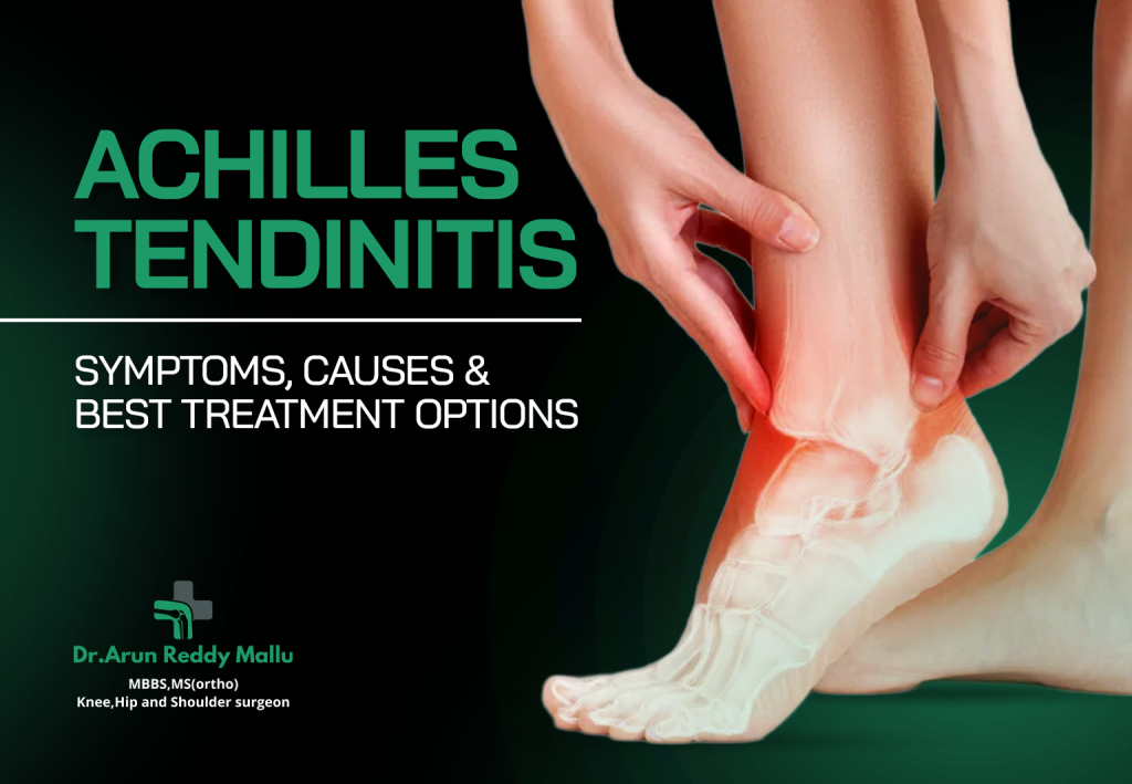
Achilles Tendinitis: Symptoms, Causes & Best Treatment Options The human body is a complex system of tissues, muscles, and tendons working in harmony to help us perform daily movements. Among these components, the Achilles tendon is one of the most critical — and yet, one of the most commonly injured. If you’ve ever experienced pain or stiffness in the back of your heel after exercise, you might be familiar with Achilles tendinitis. Achilles tendinitis is a condition characterized by inflammation of the Achilles tendon — the band of tissue that connects your calf muscles to your heel bone. This condition can affect both athletes and non-athletes alike and can range from mild irritation to debilitating pain that hinders mobility. In this article, we’ll explore the symptoms, causes, and most effective treatment options for Achilles tendinitis, enabling you to better understand and manage this widespread condition. What Is the Achilles Tendon? The Achilles tendon is the largest and strongest tendon in the human body. It plays an essential role in walking, running, jumping, and standing on tiptoe. When you push off the ground during movement, the Achilles tendon transmits force from your calf muscles to your heel, enabling you to propel yourself forward. Despite its strength, the Achilles tendon has a relatively limited blood supply, which makes it susceptible to irritation, strain, and inflammation, particularly when it is overused or stressed. Symptoms of Achilles Tendinitis Achilles tendinitis usually develops gradually rather than suddenly. Symptoms vary in severity and often worsen with continued activity. Common signs include: Pain and Stiffness Pain at the back of the heel or along the tendon, especially in the morning. Stiffness that eases as you warm up but worsens again after activity. Swelling Mild to moderate swelling around the Achilles tendon. Tenderness when touched. Limited Range of Motion Difficulty flexing the foot. Trouble walking uphill or climbing stairs. Thickening of the Tendon In some cases, the tendon may appear thicker due to chronic irritation and inflammation. Crepitus A crackling or crackling sensation may be felt around the tendon during movement. If left untreated, Achilles tendinitis can worsen and lead to more serious complications like tendon rupture, making early detection and treatment critical. Causes and Risk Factors Achilles tendinitis typically arises due to excessive strain on the tendon. The causes can be multifactorial, often involving repetitive stress and biomechanical issues. Overuse This is the most common cause: Increasing training intensity or duration too quickly. Running or jumping sports that put repetitive stress on the calf muscles. Lack of Proper Warm-Up Not adequately stretching or warming up before exercise can increase the risk of tendon stress and microscopic tears. Tight or Weak Muscles Tight calf muscles or weak supporting muscles can increase the load on the Achilles tendon during activity. Improper Footwear Poorly fitting shoes or shoes with inadequate support can alter foot mechanics and increase strain on the tendon. Biomechanical Issues Flat feet, high arches, or abnormal walking patterns can place additional stress on the tendon. Age and Activity Level Achilles tendinitis is particularly common in: Middle-aged recreational athletes. Individuals who exercise only intermittently. Sudden Changes in Activity Jumping into intensive exercise after a long period of inactivity. Understanding these causes can help you take preventive measures and make lifestyle adjustments to protect your tendon health. How Is Achilles Tendinitis Diagnosed? Diagnosis typically involves a combination of: Clinical Evaluation A healthcare provider will assess: Pain location Tenderness Swelling Range of motion Gait mechanics Physical Tests Specific movements and resistance tests can help identify tendon involvement. Imaging Studies Ultrasound:To assess inflammation and tendon structure. MRI:Used in more complex or persistent cases to rule out tears or degeneration. An accurate diagnosis ensures that you receive the most appropriate treatment and avoid worsening the condition. Best Treatment Options Achilles tendinitis can usually be managed effectively with the right treatment approach, depending on the severity of symptoms. Rest and Activity Modification Reducing or temporarily stopping activities that trigger pain is one of the most effective initial treatments. Avoid high-impact sports and replace them with low-impact activities like swimming or cycling. Ice Therapy Applying ice to the affected area for 15–20 minutes several times a day helps reduce pain and inflammation. Non-Steroidal Anti-Inflammatory Drugs (NSAIDs) Pain relievers like ibuprofen or naproxen may be recommended to reduce pain and swelling. Always consult a healthcare provider before use. Stretching and Strengthening Calf stretches and eccentric strengthening exercises are often prescribed because they: Improve flexibility Increase tendon resilience Promote healing Examples include: Wall calf stretches Heel drops on a step Resistance band exercises Physical Therapy A physical therapist can tailor a program combining: Strengthening Flexibility Balance exercises Gait correction This approach improves mechanics and reduces tendon strain. Orthotic Support Custom or over-the-counter arch supports and heel lifts can help correct biomechanical issues and decrease tendon stress. Shockwave Therapy In chronic and persistent cases, extracorporeal shockwave therapy (ESWT) may be used to stimulate healing in the tendon. Platelet-Rich Plasma (PRP) Injections PRP injections involve using concentrated platelets from your own blood to promote tendon healing. This option is typically reserved for chronic or refractory cases. Surgery Surgery is rarely required but may be considered if: Conservative treatments fail after 6–12 months There is a partial tear There’s significant degeneration (tendinosis) Surgical options may include tendon debridement, repair, or removal of scar tissue to restore healthy tendon function. Rehabilitation and Recovery Time Recovery from Achilles tendinitis varies based on severity and how early treatment begins. Mild cases may improve within a few weeks with conservative care, while more chronic cases may require several months of therapy. General recovery principles include: Gradual return to activity Ongoing stretching and strengthening Avoiding sudden increases in exercise intensity Patience and a structured rehabilitation plan significantly improve long-term outcomes. Prevention Tips Preventing Achilles tendinitis is possible with simple adjustments: Warm Up Properly Always stretch and warm up before exercising. Increase Activity Gradually Follow the 10% rule — avoid increasing training volume by more than 10% per week. Strength and Flexibility Work Maintain strong calf and
Best Exercises for Plantar Fasciitis – Expert Guide by Dr. Arun Reddy Mallu
Best Exercises for Plantar Fasciitis – Expert Guide by Dr. Arun Reddy Mallu Plantar fasciitis is one of the most common causes of heel pain, affecting athletes and non-athletes alike. This condition results when the plantar fascia — the thick band of tissue that runs along the bottom of the foot from heel to toes — becomes inflamed due to repetitive strain. While many treatments exist, one of the most effective non-surgical approaches is a tailored exercise program designed to reduce pain, improve flexibility, and strengthen the muscles supporting the arch. In this expert guide, Dr. Arun Reddy Mallu, a specialist in musculoskeletal health, explains the best exercises for plantar fasciitis that promote healing and help prevent recurrence. Whether you’re just beginning your recovery or looking to enhance your current regimen, these exercises offer a structured and evidence-based path to relief. Understanding Plantar Fasciitis Before diving into exercises, it’s important to understand the mechanics of plantar fasciitis: What causes it?Repetitive stress on the plantar fascia — often from running, long periods of standing, improper footwear, or tight calf muscles. Key symptoms:Sharp heel pain, especially with the first steps in the morning or after long rest periods, and tenderness along the arch of the foot. Why exercises help:Targeted stretching and strengthening improve flexibility, reduce strain on the fascia, and support better foot biomechanics. As part of a comprehensive treatment strategy, the right exercises can significantly decrease pain and accelerate recovery. Principles of a Good Plantar Fasciitis Exercise Program The best exercise plan for fasciitis follows three core principles: Stretching:Improve flexibility in the calf muscles and plantar fascia. Strengthening:Build support in the foot and lower leg. Mobility:Enhance range of motion in the ankle and foot joints. These components together decrease stress on the plantar fascia and improve gait mechanics. Top 12 Exercises for Plantar Fasciitis Relief Below are the best exercises recommended by experts — including Dr. Arun Reddy Mallu — designed to combat inflammation, strengthen supporting muscles, and improve flexibility. Calf Stretch Against the Wall Purpose: Loosens the tight calf muscles (gastrocnemius and soleus) that contribute to plantar fascia strain. How to do it: Stand facing a wall. Place one foot behind the other. Keep the backleg straight with heel on the ground. Lean forward slightly until you feel a stretch in the calf. Hold for 30 seconds, repeat 3 times on each leg. Tip: Keep toes pointing straight ahead to ensure proper muscle engagement. Towel Stretch Purpose: Targets the plantar fascia and calf muscles. How to do it: Sit on the floor with legs straight. Loop a towel around the ball of your foot. Gently pull the towel toward you. Hold for 30 seconds; repeat 3 times per foot. This is particularly effective first thing in the morning. Plantar Fascia Stretch Purpose: Specifically stretches the plantar fascia for targeted relief. How to do it: Sit in a chair. Cross one foot over the oppositeknee. Hold the base of your toes and pull them backward until you feel a stretch in your arch. Hold for 15–30 seconds; repeat 3 times per foot. Rolling Stretch with a Ball Purpose: Massage-like relief; increases blood flow to the fascia. How to do it: Sit and place a tennis ball, golf ball, or frozen water bottle under your foot. Roll the arch of your foot over the object slowly. Do this for 2–3 minutes per foot. Pro tip: Use a frozen bottle to help reduce inflammation. Toe Curls with Towel Purpose: Strengthens the small muscles in the foot and arch stabilization. How to do it: Place a small towel on the floor. Use your toes to scrunch the towel toward you. Repeat 10–15 times per foot. Big Toe Stretch Purpose: Enhances flexibility and range of motion in the big toe — which affects arch mechanics. How to do it: Sit with feet flat on the floor. Lift your big toe while keeping other toes grounded. Hold for 5 seconds and repeat 10 times. Ankle Circles Purpose: Improves ankle mobility and neuromuscular coordination. How to do it: Sit with your leg extended. Rotate your ankle in slow circles — 10 clockwise, then 10 counter-clockwise. Repeat 3 sets per foot. Heel Raises Purpose: Strengthens the calf muscles and supports foot biomechanics. How to do it: Stand with feet hip-width apart. Rise up on your toes, lifting heels off the floor. Slowly lower back down. Do 2–3 sets of 10–15 reps. Progression: Perform on a step to increase range of motion. Resistance Band Foot Flexion Purpose: Strengthens the muscles that support the arch. How to do it: Sit with a resistance band looped around the ball of your foot. Flex your foot against the resistance. Do 3 sets of 10–15 reps. This can be done daily with a light band. Walking on Sand (If Available) Purpose: Natural strengthening and stretch for foot muscles. How to do it: Spend 10–15 minutes walking barefoot on sand. It forces active engagement of the foot muscles and provides gentle stretch. Achilles Tendon Stretch Purpose: Targets the Achilles tendon, which shares load with the plantar fascia. How to do it: Stand facing a wall. Step one foot back and bend both knees. Press the back heel down. Hold for 30 seconds; repeat 3 times per side. Arch Lifts (Doming Exercise) Purpose: Strengthens intrinsic foot muscles that maintain the arch. How to do it: Stand or sit with feet flat. Lift the arch without curling the toes. Hold 3–5 seconds; repeat 10–15 times. Consistency matters more than intensity. When and How Often to Do These Exercises For optimal results: Daily routine:Stretching exercises (1–4) should be done every morning and evening. Strengthening:Perform exercises (5–12) once per day or every other day. Listen to your body:Some discomfort is normal, but sharp pain isn’t — adjust intensity accordingly. Warm up first:A light 5-minute walk before exercises helps prepare the tissues. Those in early recovery stages may need to reduce intensity and focus more on gentle stretches initially. Tips for Best Results ✅ Wear supportive footwear — avoid walking barefoot on hard surfaces✅ Avoid high-impact sports until pain subsides✅ Ice the heel for 10–15 minutes after workouts✅ Combine exercises with shock-absorbing insoles or orthotics✅ Maintain
Muscle Cramps: What You Should Know
Muscle Cramps: What You Should Know Muscle cramps are sudden, involuntary contractions of one or more muscles that can cause sharp pain and temporary discomfort. They can occur during physical activity, while resting, or even in the middle of the night. Almost everyone experiences muscle cramps at some point in life, yet many people are unsure why they happen or how to prevent them effectively. Although most muscle cramps are harmless and short-lived, frequent or severe cramps may interfere with daily activities and sleep, and in some cases, signal an underlying medical issue. Understanding the causes, symptoms, treatment options, and preventive strategies can help you manage muscle cramps more effectively and improve your overall muscle health. What Are Muscle Cramps? A muscle cramp is a sudden and uncontrollable tightening of a muscle or muscle group. The affected muscle may feel hard or knotted and can cause significant pain until it relaxes. Muscle cramps most commonly affect the legs—especially the calves—but they can also occur in the thighs, feet, hands, arms, abdomen, and even the chest. Cramps typically last from a few seconds to several minutes. After the cramp subsides, the muscle may remain sore or tender for hours. While occasional cramps are usually not a cause for concern, recurring episodes should not be ignored. Common Causes of Muscle Cramps Muscle cramps can develop for many reasons, often related to muscle strain, hydration levels, or nerve function. Some of the most common causes include: Muscle Overuse and Fatigue Overworking muscles during exercise or physical labor can lead to cramps, especially when the body is not conditioned for the activity. Repetitive movements, prolonged standing, or sudden increases in activity levels can strain muscles and trigger painful spasms. Dehydration Inadequate fluid intake is one of the most common causes of muscle cramps. When the body loses fluids through sweating—especially in hot weather or during intense exercise—muscles may not function properly, increasing the risk of cramping. Electrolyte Imbalance Electrolytes such as potassium, magnesium, calcium, and sodium play a vital role in muscle contraction and relaxation. Low levels of these minerals can disrupt muscle function and lead to cramps. Poor nutrition, excessive sweating, and certain medical conditions can contribute to electrolyte imbalance. Poor Blood Circulation Reduced blood flow to muscles can cause cramping, particularly during physical activity. This type of cramp often improves with rest and may indicate circulation-related issues that require medical evaluation. Nerve Compression Pressure on nerves—especially in the spine—can result in muscle cramps. This may occur due to posture issues, spinal conditions, or prolonged sitting. Cramps related to nerve compression may worsen with walking or standing for long periods. Medical Conditions and Medications Certain health conditions such as diabetes, thyroid disorders, kidney disease, and neurological conditions may increase the likelihood of muscle cramps. Some medications, including diuretics and cholesterol-lowering drugs, can also affect muscle function or electrolyte balance. Age-Related Changes As people age, muscle mass naturally decreases, and nerves may become more sensitive. This makes older adults more prone to muscle cramps, particularly at night. Symptoms of Muscle Cramps Muscle cramps are usually easy to recognize. Common symptoms include: Sudden, sharp pain in a muscle Tightening or hardening of the muscle Visible muscle twitching or knotting Difficulty moving the affected area Lingering soreness after the cramp subsides Night-time leg cramps, also known as nocturnal cramps, are especially common and can disrupt sleep, leading to fatigue and reduced quality of life. When Should You See a Doctor? Occasional muscle cramps are usually harmless. However, medical attention is recommended if cramps: Occur frequently or worsen over time Are severe or long-lasting Interfere with daily activities or sleep Are accompanied by muscle weakness, swelling, or numbness Occur without obvious triggers such as exercise or dehydration Persistent cramps may indicate an underlying medical condition that requires proper diagnosis and treatment. How Are Muscle Cramps Diagnosed? Diagnosis usually begins with a detailed medical history and physical examination. Your doctor may ask about: Frequency and duration of cramps Activities or situations that trigger cramps Diet, hydration habits, and exercise routine Medications and existing health conditions In some cases, blood tests may be recommended to check electrolyte levels, kidney function, or hormone balance. Additional tests may be ordered if nerve or circulation problems are suspected. Treatment Options for Muscle Cramps Most muscle cramps can be relieved with simple self-care measures. Treatment focuses on relaxing the affected muscle and preventing recurrence. Stretching and Massage Gently stretching the cramped muscle can help relieve pain and stop the spasm. Massaging the area improves blood flow and promotes muscle relaxation. Heat and Cold Therapy Applying heat helps relax tight muscles, while cold therapy can reduce soreness and inflammation after the cramp has resolved. Proper Hydration Drinking enough water throughout the day is essential, especially during physical activity or hot weather. Adequate hydration supports normal muscle function. Balanced Nutrition Including foods rich in potassium, magnesium, and calcium—such as fruits, vegetables, dairy products, nuts, and whole grains—supports healthy muscle contractions. Medications (If Needed) In cases where cramps are frequent or related to an underlying condition, medications may be prescribed to manage symptoms or correct imbalances. These should only be taken under medical supervision. Preventing Muscle Cramps Preventive strategies can significantly reduce the frequency of muscle cramps: Stretch regularly, especially before and after exercise Warm up muscles before intense physical activity Maintain proper hydration throughout the day Eat a nutrient-rich, balanced diet Avoid prolonged sitting or standing in one position Maintain good posture and ergonomic habits Address underlying medical conditions promptly For individuals prone to night-time cramps, gentle stretching before bedtime can be particularly helpful. Conclusion Muscle cramps are a common condition that can affect anyone, regardless of age or activity level. While they are often temporary and harmless, frequent or severe cramps should not be ignored. Understanding the causes—such as dehydration, muscle fatigue, electrolyte imbalance, and medical conditions—can help in managing and preventing these painful episodes. If muscle cramps persist or significantly impact your quality of life, expert
How to Prepare for Knee & Shoulder Surgery This Winter in Hyderabad
How to Prepare for Knee & Shoulder Surgery This Winter in Hyderabad Knee and shoulder problems can significantly affect your quality of life, limiting mobility, independence, and daily comfort. When conservative treatments such as medication, physiotherapy, or injections no longer provide relief, surgery may become the most effective solution. For many patients in Hyderabad, winter is an ideal season to undergo knee or shoulder surgery, thanks to cooler temperatures and better post-operative comfort. However, successful surgery is not just about the procedure itself—it depends heavily on how well you prepare beforehand. From physical conditioning and medical readiness to home planning and mental preparation, every step matters. This guide will help you understand how to prepare effectively for knee and shoulder surgery this winter in Hyderabad. Understand Your Condition and Surgical Procedure Preparation begins with awareness. Before scheduling surgery, take time to understand your diagnosis clearly. Knee and shoulder surgeries can vary widely, including procedures such as: Total orpartial knee replacement Arthroscopic knee surgery Shoulder arthroscopy Rotator cuff repair Shoulder replacement Your orthopedic surgeon will explain the nature of your joint problem, why surgery is required, and which procedure is most suitable. Understanding the expected benefits, possible risks, and recovery timeline helps you set realistic expectations and reduces anxiety. Do not hesitate to ask questions about pain control, hospital stay duration, mobility after surgery, and rehabilitation requirements. A well-informed patient is better prepared both physically and mentally. Complete Pre-Surgical Medical Evaluation Before surgery, you will undergo a comprehensive medical evaluation to ensure your body is ready for anesthesia and healing. This typically includes: Blood tests Heart evaluation (ECG) Imaging studies such as X-rays or MRI General health assessment If you have conditions such as diabetes, high blood pressure, asthma, or heart disease, they must be well controlled before surgery. Inform your doctor about all medications, supplements, or herbal remedies you take, as some may need to be paused before surgery to reduce bleeding risks or anesthesia complications. Strengthen Your Body Before Surgery (Prehabilitation) One of the most important yet often overlooked steps is prehabilitation, or preparing your muscles before surgery. For Knee Surgery Strengthening the thigh, calf, and hip muscles helps support the knee joint. Gentle exercises improve flexibility and reduce stiffness after surgery. Low-impact activities such as walking or cycling are beneficial. For Shoulder Surgery Maintaining shoulder range of motion within pain limits is important. Strengthening surrounding muscles supports faster recovery. A physiotherapist can guide you with safe pre-operative exercises. Patients who engage in prehabilitation often experience less post-operative pain, quicker mobility, and better long-term outcomes. Focus on Nutrition and Hydration Your body needs proper fuel to heal efficiently. In the weeks leading up to surgery: Increase protein intake to support tissue repair. Eat fresh fruits and vegetables for vitamins and antioxidants. Ensure adequate calcium and vitamin D for bone health. Stay well hydrated to maintain circulation and joint lubrication. Avoid crash diets or skipping meals before surgery. Good nutrition plays a vital role in reducing infection risk and promoting faster recovery. Adjust Medications, Smoking, and Alcohol Habits Certain lifestyle habits can negatively affect surgery and healing. Smokingreduces blood flow and delays wound healing. Quitting even a few weeks before surgery can significantly improve outcomes. Alcohol consumptionshould be minimized or stopped prior to surgery, as it can interfere with anesthesia and increase bleeding risk. Follow your doctor’s advice regarding medications, especially blood thinners and pain relievers. These adjustments may feel challenging, but they directly impact your surgical success. Prepare Your Home for Recovery After knee or shoulder surgery, simple daily tasks can become temporarily difficult. Preparing your home in advance ensures safety and comfort. Safety Modifications Remove loose rugs and clutter to prevent falls. Ensure adequate lighting, especially at night. Install grab bars in bathrooms if needed. Comfort Setup Arrange a recovery area with pillows, blankets, water, medicines, and essentials within easy reach. Keep frequently used items at waist level to avoid bending or reaching. Use chairs with armrests to assist sitting and standing. Support System Arrange for a family member or caregiver to help with cooking, bathing, and errands during the first few days. Prepare meals in advance to reduce daily strain. Plan Your Hospital Stay Knowing what to expect during your hospital stay can ease stress. Pack essentials such as: Comfortable, loose clothing Slip-resistant footwear Personal toiletries Medical documents and identification Mobile phone and charger Also, arrange transportation in advance, as driving is not allowed immediately after surgery. Winter-Specific Considerations Winter offers certain advantages for joint surgery: Cooler temperatures can reduce swelling and discomfort. Patients often sleep better and experience less fatigue. Rehabilitation exercises are more manageable without excessive heat. However, take precautions when moving outdoors, as slippery surfaces can increase fall risk. Wearing appropriate footwear and avoiding unnecessary outdoor activity during early recovery is essential. Mental and Emotional Preparation Surgery can cause emotional stress, especially for first-time patients. Preparing mentally is just as important as physical readiness. Practice relaxation techniques such as deep breathing or meditation. Maintain a positive outlook and trust the recovery process. Set realistic goals and understand that healing takes time. Staying mentally calm and optimistic helps your body respond better to treatment and rehabilitation. Follow Pre-Operative Instructions Carefully Your surgeon will provide detailed instructions before surgery, including: When to stop eating and drinking Which medications to take or avoid Personal hygiene recommendations Following these instructions strictly helps prevent complications and ensures your surgery proceeds as planned. Conclusion Preparing for knee or shoulder surgery is a journey that involves thoughtful planning, physical readiness, and emotional strength. From understanding your condition and strengthening your body to preparing your home and mindset, each step plays a vital role in ensuring a smooth surgical experience and faster recovery. If you are considering knee or shoulder surgery this winter in Hyderabad, expert guidance makes all the difference. Dr. Arun Reddy Mallu at Continental Hospitals is renowned for his expertise in orthopedic care and joint surgeries. With a
Frozen Shoulder (Adhesive Capsulitis): Early Signs, Stages & Recovery Tips
Frozen Shoulder (Adhesive Capsulitis): Early Signs, Stages & Recovery Tips Frozen shoulder, medically known as adhesive capsulitis, is a painful and often debilitating condition of the shoulder joint. It can gradually restrict your ability to move your arm comfortably — affecting daily activities like dressing, reaching overhead, or even sleeping. While many people shrug off shoulder stiffness as “just getting older” or a consequence of minor injury, frozen shoulder is a specific clinical entity that benefits from early recognition and targeted management. In this blog, we’ll walk you through: What frozen shoulder really is Early warning signs The four classic stages of the condition Practical recovery tips When to seek expert care, including insights from Arun Reddy Malluat Continental Hospitals What Is Frozen Shoulder? Frozen shoulder refers to inflammation and thickening of the capsule of the shoulder joint. The capsule is a connective tissue envelope that surrounds the shoulder, providing support and stability. In adhesive capsulitis, this capsule becomes inflamed, tight, and less elastic — effectively “freezing” the shoulder’s range of motion. Although the exact cause isn’t always clear, it’s most commonly associated with: Long periods of shoulder immobility (after injury or surgery) Diabetes (highly correlated) Thyroid disorders Prior shoulder trauma Unlike acute injuries or arthritis, frozen shoulder evolves gradually and can take months to resolve. Early Signs & Symptoms To Watch For Recognizing frozen shoulder early improves the chances of effective treatment and faster recovery. Here are the initial warning signs: Gradual Onset of Pain The first symptom is usually a dull or aching pain deep within the shoulder. It may be worse at night or with specific movements. Often, people mistake this for muscle strain or fatigue. Progressive Stiffness Within weeks, your shoulder may feel less flexible. You might notice difficulty with movements like: Reaching above your head Reaching behind your back Rotating your arm outward This stiffness is typically painless at first but progresses. Pain With Activity Simple tasks such as combing hair, putting on a jacket, or reaching into a cupboard can start to hurt. This is often the first time many patients realize something more than “simple muscle tightness” is happening. Sleep Disturbances Persistent shoulder pain can interrupt sleep — especially when lying on the affected side. Difficulty achieving comfortable sleep positions is a common early complaint. Reduced Range of Motion Your shoulder simply won’t move as freely as before. Clinically, both active movement (you move) and passive movement (someone else moves your arm) become restricted. If you notice stiffness that doesn’t improve over several weeks — especially accompanied by worsening pain — it’s worth consulting a specialist. Four Stages of Frozen Shoulder Frozen shoulder doesn’t happen overnight. It evolves through four well-described stages. Each stage has distinctive features and implications for management. Stage 1 — The “Freezing” Stage Duration: 6–9 months Symptoms: Gradual increase in pain Stiffness starts, particularly with rotation Sleep disturbances During this stage, shoulder movement becomes increasingly restricted as inflammation of the joint capsule progresses. Pain is the dominant symptom. Many people seek help at this point, but it’s often mistaken for tendonitis or bursitis. Stage 2 — The “Frozen” Stage Duration: 4–12 months Symptoms: Significant stiffness Pain may decrease slightly Range of motion is severely limited In this stage, the shoulder becomes very tight. Although pain might lessen, stiffness has worsened — and daily activities become harder. Tasks such as fastening a bra, reaching overhead, or tucking in a shirt become difficult. Stage 3 — The “Thawing” Stage Duration: 6–12 months Symptoms: Gradual improvement in motion Less pain Function slowly returns The body begins to “remodel” the stiffened joint capsule. Daily activities become easier, although full motion may not be restored until later. Stage 4 — Recovery & Remodeling Duration: Variable (weeks to months after thawing) Symptoms: Increasing flexibility Minimal pain Functional improvement This stage represents recovery — improvements continue, although the timeline varies widely among individuals. Managing Frozen Shoulder: Practical Tips Effective management depends on early recognition and appropriate exercises, often combined with medical guidance. Here’s a comprehensive plan to support recovery. Gentle, Consistent Stretching The cornerstone of recovery is controlled, gentle stretching to maintain and gradually increase range of motion: Pendulum stretch:Let your arm hang and gently swing in small circles. Towel stretch:Grasp a towel behind your back with both hands and gently pull. Cross-body reach:Use your good arm to lift the affected arm across your body. Key: Never force movement to the point of sharp pain. Heat & Cold Therapy Heatbefore stretching can ease stiffness and relax muscles. Coldafter activity can reduce inflammation and discomfort. Use moist heat or warm showers before exercises for best effect. Pain Management Over-the-counter NSAIDs (e.g., ibuprofen) can help with inflammation and pain. Always consult a healthcare provider before starting medications, especially if you have gastric or kidney issues. Professional Physical Therapy A trained physiotherapist can tailor exercises and manual therapy to your condition — crucial for optimal recovery. Techniques may include: Passive stretching Mobilization exercises Strengthening routines once motion improves Consistency is key — daily therapy can significantly improve outcomes. Injections & Interventions For cases where pain severely limits function, a doctor may recommend: Corticosteroid injectionsto reduce inflammation Nerve blocksfor pain relief Hydrodilatation(capsular distension with saline/contrast) These should be guided by an experienced specialist. Avoid Prolonged Immobilization Keeping your shoulder immobile (like in a sling) for long periods can worsen stiffness. Balance rest with gentle motion. When To See a Specialist If stiffness and pain persist for more than 6–8 weeks, or if everyday activities are compromised, getting expert evaluation is important. At Continental Hospitals, orthopedic specialists — including Dr. Arun Reddy Mallu, who has extensive experience in shoulder conditions and minimally invasive therapies — can provide: Accurate diagnosis (clinical + imaging) Personalized treatment plans Guidance through physical therapy Advanced interventional options if needed Dr. Arun Reddy Mallu emphasizes early recognition and tailored therapy to prevent long-term disability and optimize recovery. Myths & Misconceptions Let’s debunk a few common myths about frozen shoulder: It only happens to older people. While more
What Are the Best Indications for Seeing a Knee and Shoulder Surgeon in Hyderabad?

What Are the Best Indications for Seeing a Knee and Shoulder Surgeon in Hyderabad? The knee and shoulder are two of the most complex and heavily used joints in the human body. From walking and climbing stairs to lifting, throwing, and performing daily activities, these joints play a crucial role in maintaining mobility and independence. When pain, stiffness, or injury affects them, daily life can quickly become uncomfortable and restricted. Many people delay seeing a specialist, assuming joint pain will resolve on its own. However, knowing when to consult a knee and shoulder surgeon can prevent long-term damage and improve recovery outcomes. This blog explains the key indications for seeking expert orthopedic care and highlights the importance of consulting an experienced specialist in Hyderabad. Who Is a Knee and Shoulder Surgeon? A knee and shoulder surgeon is an orthopedic specialist trained to diagnose and treat conditions affecting these joints. Their expertise includes both non-surgical management and advanced surgical procedures, such as arthroscopy, ligament reconstruction, cartilage repair, and joint replacement. These specialists handle conditions caused by: Sports injuries Accidents or trauma Degenerative diseases like arthritis Overuse and repetitive strain Age-related joint wear Seeing the right specialist at the right time can significantly reduce pain, restore movement, and prevent complications. Persistent Knee or Shoulder Pain One of the most common indications to see a knee and shoulder surgeon is pain that does not improve with rest or basic treatment. Occasional soreness after activity is normal. However, you should seek expert care if: Pain lasts more than a few weeks Pain increases with movement or weight-bearing Pain occurs even during rest or sleep Persistent pain may indicate ligament injuries, tendon damage, cartilage wear, or early arthritis. Ignoring ongoing pain can lead to worsening joint damage and reduced mobility. Limited Range of Motion Difficulty moving your knee or shoulder through its normal range is a strong warning sign. This may include: Trouble bending or straightening the knee Inability to lift the arm overhead Stiffness that does not improve with exercises Conditions such as frozen shoulder, rotator cuff tears, meniscus injuries, or cartilage degeneration often cause restricted movement. Early evaluation by a specialist helps restore function before stiffness becomes permanent. Swelling, Warmth, or Inflammation Swelling around the knee or shoulder, especially when accompanied by pain or warmth, suggests internal joint irritation or injury. This may result from: Ligament or meniscus tears Inflammation of tendons or bursae Joint fluid accumulation Infection or inflammatory arthritis Persistent swelling should never be ignored, as it can lead to long-term joint damage if untreated. Joint Instability or “Giving Way” If your knee feels unstable or your shoulder feels like it might slip out of place, it may indicate ligament damage or joint instability. Common causes include: ACLor PCL injuries in the knee Shoulder dislocations or labral tears Weak supporting structures around the joint Instability increases the risk of repeated injuries and cartilage damage. A knee and shoulder surgeon can assess the joint and recommend appropriate treatment, including surgical stabilization if needed. Sudden Injury or Trauma Any significant injury caused by a fall, accident, or sports activity should be evaluated by an orthopedic specialist, especially if it results in: Immediate pain and swelling Inability to move the joint Deformity or abnormal alignment Prompt assessment ensures accurate diagnosis and timely treatment, which is critical for proper healing and long-term joint health. Difficulty Performing Daily Activities When knee or shoulder problems interfere with everyday tasks, it is a clear indication to seek medical attention. These difficulties may include: Pain while walking, standing, or climbing stairs Trouble lifting objects or reaching overhead Difficulty getting in and out of chairs or beds Such limitations often indicate progressive joint problems that require professional care to prevent further decline. Chronic Arthritis and Degenerative Joint Conditions Arthritis is a common cause of knee and shoulder pain, especially with advancing age. While early arthritis can often be managed conservatively, you should consult a knee and shoulder surgeon if: Pain is persistent and worsening Joint stiffness limits daily function Medications and physiotherapy no longer provide relief A specialist can guide you through advanced treatment options, including injections or surgical solutions, when appropriate. Sports Injuries That Do Not Heal Athletes and physically active individuals frequently experience knee and shoulder injuries. While many minor injuries heal with rest and rehabilitation, specialist evaluation is required if: Pain persists despite physiotherapy There is repeated swelling or instability Performance and strength do not return Conditions such as ligament tears, cartilage injuries, and tendon ruptures often require surgical intervention for full recovery. Recurrent Dislocations or Repeated Injuries If your shoulder dislocates repeatedly or your knee suffers frequent injuries, it usually indicates an underlying structural problem. Recurrent injuries can weaken the joint further and accelerate arthritis. A knee and shoulder surgeon can identify the cause and provide long-term solutions to restore stability and prevent recurrence. Failure of Previous Treatment If you have already tried: Medications Physiotherapy Injections Previous surgery and still experience pain or limited function, it is important to consult a specialist with advanced expertise. Revision surgeries and advanced procedures can often resolve unresolved joint problems. Expert Knee and Shoulder Care in Hyderabad Hyderabad is home to advanced orthopedic care, and choosing the right specialist plays a major role in treatment success. Dr. Arun Reddy Mallu, Senior Consultant Orthopedic Surgeon at Continental Hospitals, Hyderabad, is a trusted name in knee and shoulder care. With extensive experience in arthroscopy, sports injuries, trauma, and joint reconstruction, he provides comprehensive treatment tailored to each patient’s needs. Dr. Arun Reddy Mallu specializes in: Knee ligament and meniscus injuries Shoulder rotator cuff tears and instability Hip joint conditions and replacement surgeries Arthroscopic (minimally invasive) procedures Sports injury management Robotic-assisted joint replacement surgeries Joint preservation and replacement surgeries At Continental Hospitals, patients benefit from modern diagnostic tools, advanced surgical technology, and a multidisciplinary rehabilitation approach — ensuring safe treatment and faster recovery. Conclusion Knee and shoulder problems should never be ignored, especially when pain,