
Warning Signs of Infection After Knee Replacement Surgery
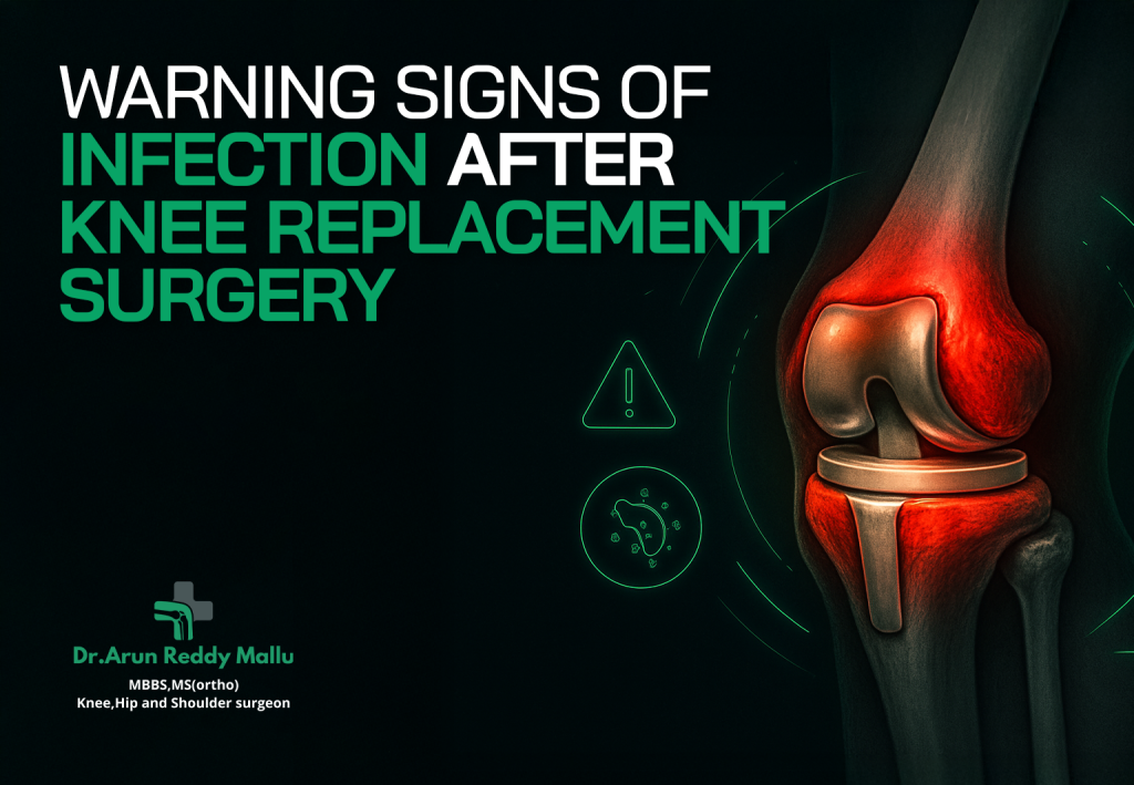
Warning Signs of Infection After Knee Replacement Surgery Knee replacement surgery, also known as knee arthroplasty, is one of the most commonly performed orthopedic surgeries today, offering relief to millions suffering from arthritis, chronic pain, or knee injuries. While the procedure itself has a high success rate, one of the major concerns after knee replacement surgery is the risk of infection. Infection can complicate recovery and may lead to serious health issues if not detected and treated early. In this blog post, we will discuss the warning signs of infection after knee replacement surgery, the factors that increase the risk of infection, and the necessary steps to take if you suspect an infection. We’ll also highlight the expertise of Dr. Arun Reddy Mallu at Continental Hospital, a renowned orthopedic specialist, who can help you navigate post-surgery recovery safely and effectively. Why Is Infection a Concern After Knee Replacement Surgery? Infections following knee replacement surgeries can range from mild to severe and can occur at different stages of the healing process. An infection might affect the incision site (superficial infection) or, in more serious cases, the artificial joint (deep infection), which can be difficult to treat and might require additional surgery. The risk of infection is always present after surgery, but understanding the early warning signs can significantly improve your chances of successful treatment. The body typically fights infections with its immune system, but surgical interventions—like knee replacements—can introduce bacteria into the body. This can happen if proper sterile techniques aren’t followed during surgery, if the incision site is exposed to germs during the recovery period, or if the patient’s immune system is compromised in any way. Early Warning Signs of Infection After Knee Replacement Surgery Redness and Swelling Around the Surgical Site After knee replacement surgery, some swelling is expected as part of the normal healing process. However, if the swelling worsens or the skin around the incision turns red or hot to the touch, it could indicate an infection. This is often a sign that the body is trying to fight an infection and that the area is inflamed. You should contact your healthcare provider immediately if you notice these symptoms. Fever or Chills A low-grade fever (below 101°F) is not uncommon following surgery, as it’s part of the body’s natural healing response. However, a high fever (above 101°F), or fever that persists for more than a few days, could be a sign of a systemic infection. Chills often accompany fever, and together they may indicate that an infection is present in the body. Increased Pain or Tenderness While some discomfort is expected after knee replacement surgery, a sudden increase in pain or unusual tenderness around the incision site can be a warning sign. If the pain becomes more severe and is not alleviated by prescribed pain medications, or if it’s associated with any other symptoms like fever or redness, it could suggest an infection. Pus or Drainage from the Incision Any discharge from the surgical site that is green, yellow, or has a foul odor is a clear sign of infection. Pus is produced by the body when it tries to fight an infection, and it often indicates the presence of bacteria. If you notice any drainage or secretion from your incision, it’s essential to seek medical attention promptly to prevent the infection from spreading. Limited Range of Motion After knee replacement surgery, patients are encouraged to begin physical therapy to restore mobility. However, if you experience a sudden loss of range of motion or an inability to move the knee joint, especially along with other symptoms like fever or swelling, it could indicate an infection. The infection can cause stiffness and limit the joint’s ability to function properly. Unusual Odor An unpleasant smell emanating from the surgical site is another potential warning sign of infection. When bacteria invade the surgical area, they can cause an infection that leads to an unpleasant odor as the body fights the infection. Fatigue or Malaise Post-surgery fatigue is normal, but if you feel excessively tired or unwell—especially if you’re also experiencing fever, redness, or swelling—it could be a sign that your body is fighting an infection. Malaise, a general feeling of being unwell, can often accompany an infection, so it’s important to monitor how you feel in the days following surgery. Worsening of Pre-existing Symptoms In some cases, infection after knee replacement can worsen the symptoms that prompted the surgery in the first place. If your pain, discomfort, or inability to use your knee increases or if new symptoms arise, it’s crucial to reach out to your healthcare provider. Factors That Increase the Risk of Infection While infection can happen to anyone after knee replacement surgery, several factors can increase the risk. Some of these factors include: Pre-existing Medical Conditions: Conditions like diabetes, uncontrolled diabetes, rheumatoid arthritis, or immunocompromised states (due to medications or treatments) can make it more difficult for the body to fight off infection. Obesity: Excess weight puts additional strain on the knee and can also make healing more challenging, increasing the risk of infection. Chronic Smoking:Smoking impairs blood circulation, delays wound healing, and weakens the immune system, making smokers more susceptible to infections after surgery. Poor Hygiene or Incision Care: If the surgical site isn’t properly cleaned or protected, bacteria can easily enter the incision, leading to infection. Previous Joint Infections: If you’ve had a prior infection in the joint, you may be at a higher risk for future infections after knee replacement surgery. Inadequate Post-Surgery Care: Failing to follow the post-surgery instructions carefully, such as taking prescribed antibiotics or avoiding activities that might stress the knee, can increase the likelihood of complications. What to Do If You Suspect an Infection If you suspect that you may have an infection after knee replacement surgery, it’s important to take immediate action. Here are the steps you should follow: Contact Your Surgeon: Reach out to your orthopedic surgeon or healthcare provider as soon as possible. If you’re under the care of Arun
Discoid Meniscus: What It Is and How It’s Treated
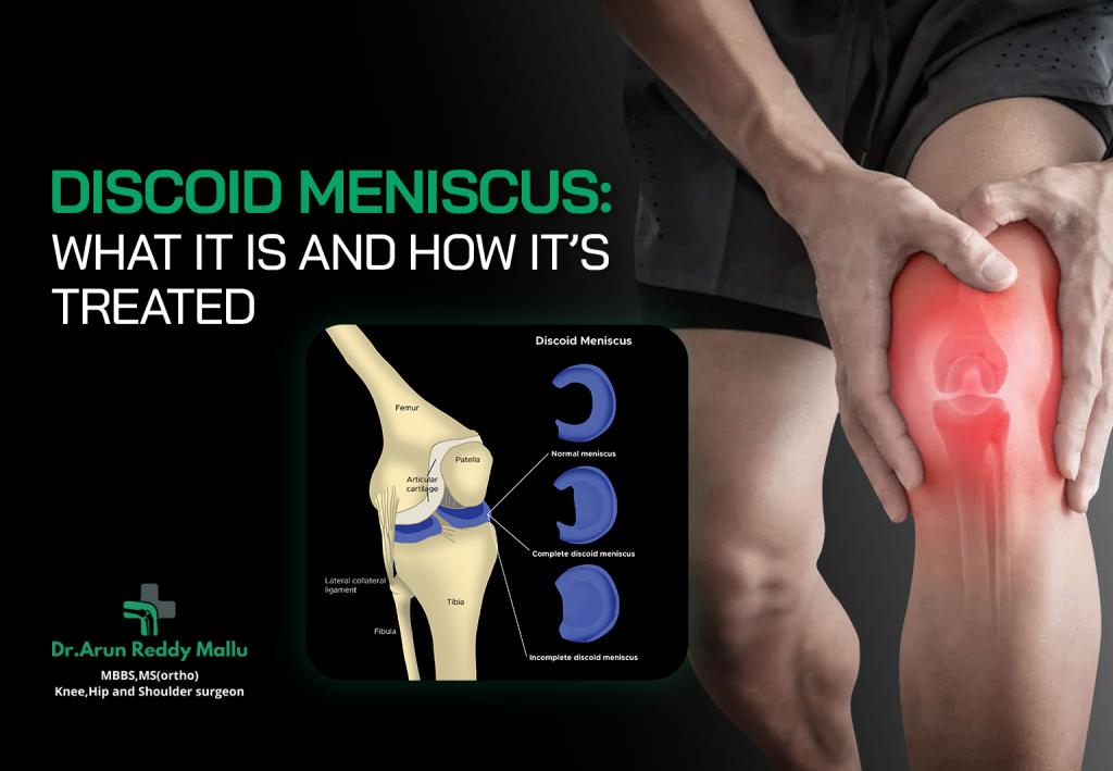
Discoid Meniscus: What It Is and How It’s Treated The knee is one of the most complex and important joints in the human body, allowing us to walk, run, squat, and jump. Inside the knee are two crescent-shaped pieces of cartilage known as the menisci — the medial (inner) and lateral (outer) meniscus. These structures act as shock absorbers between the thigh bone (femur) and the shin bone (tibia), distributing weight and providing stability. However, in some individuals, particularly children and adolescents, the meniscus may develop in an abnormal, disc-like shape rather than the typical crescent form. This condition is called a Discoid Meniscus. Though it can exist without symptoms, it often leads to pain, swelling, and mechanical issues in the knee, especially during activity. Let’s explore what a discoid meniscus is, its symptoms, causes, diagnosis, and treatment options available — including advanced care approaches offered by Dr. Arun Reddy Mallu at Continental Hospital, Hyderabad. What Is a Discoid Meniscus? A Discoid Meniscus is a congenital (present at birth) variation in which the meniscus is abnormally shaped like a disc rather than a crescent. This thicker, wider cartilage can cover a larger portion of the tibial surface than normal. The condition most commonly affects the lateral meniscus (outer part of the knee) and is more prevalent in children and young adults. In some cases, the discoid meniscus is more prone to tearing or becoming unstable because its shape and attachment to the surrounding tissues are abnormal. This makes it less flexible and more vulnerable during twisting or high-impact activities. Types of Discoid Meniscus Discoid menisci are classified into three main types based on their shape and attachment pattern: Incomplete Discoid Meniscus:The meniscus is partially disc-shaped and covers more of the tibial surface than usual but not entirely. Complete Discoid Meniscus:The meniscus completely covers the tibial surface and is thicker and more prone to tears. Wrisberg Variant (Hypermobile Meniscus):This type lacks normal posterior attachments, leading to excessive movement of the meniscus. It often causes “snapping” sensations in the knee. Causes of Discoid Meniscus The exact cause of a discoid meniscus isn’t fully understood, but it is believed to result from an abnormal development of the meniscus during fetal growth. Normally, the meniscus begins as a disc and reshapes into a crescent during early development. In cases of discoid meniscus, this reshaping process doesn’t occur completely. Although it is congenital, the condition might not present symptoms until later in childhood or adolescence when physical activity increases or a tear occurs. There is also a genetic predisposition, as discoid meniscus can sometimes run in families. Symptoms of Discoid Meniscus Many people with a discoid meniscus may not experience symptoms, and the condition is often discovered incidentally during an MRI or surgery for another knee issue. However, when symptoms do appear, they may include: Knee pain(especially during movement or twisting) Swellingor stiffness around the joint Popping or clicking sounds Locking or catching sensation Instabilityor “giving way” of the knee Limited range of motion A torn discoid meniscus can cause more severe symptoms, such as sharp pain, swelling, and an inability to fully straighten or bend the knee. How Is Discoid Meniscus Diagnosed? Early diagnosis is essential to prevent long-term damage to the knee joint. Your doctor will begin by taking a detailed medical history and performing a physical examination. During the exam, they may check for clicking, tenderness, range of motion, and joint stability. Imaging Tests: X-rays:While an X-ray doesn’t show the meniscus directly, it helps rule out bone injuries or abnormalities. MRI (Magnetic Resonance Imaging):The most effective diagnostic tool for identifying a discoid meniscus. MRI provides detailed images of the cartilage, showing its shape, thickness, and any associated tears or inflammation. Arthroscopy:In some cases, an arthroscopic procedure may be used both for diagnosis and treatment. It involves inserting a small camera into the knee jointfor a direct view of the meniscus. Treatment Options for Discoid Meniscus The treatment approach depends on several factors — such as the type of discoid meniscus, severity of symptoms, age, and level of activity. Non-Surgical Management If the discoid meniscus is asymptomatic or only mildly symptomatic, non-surgical treatments may be recommended, including: Rest and Activity Modification:Avoiding activities that cause pain or clicking. Physical Therapy:Strengthening the muscles around the knee improves stability and joint function. Medications:Over-the-counter pain relievers or anti-inflammatory drugs can help reduce swelling and discomfort. Regular Monitoring:Periodic follow-ups with imaging to ensure the condition is not worsening. Non-surgical management works best when the discoid meniscus is stable and there are no tears. Surgical Treatment If symptoms persist or if there is a meniscal tear, surgery may be required. Modern surgical techniques aim to preserve as much of the meniscus as possible, as complete removal can lead to early arthritis. The main surgical procedures include: Arthroscopic Saucerization (Meniscoplasty) This is the most common and preferred surgical method. The surgeon reshapes the discoid meniscus into a normal, crescent form, preserving its function while reducing the risk of future tears. Meniscal Repair If the meniscus has torn, the surgeon may repair it using sutures. Preserving the tissue is especially important in younger patients to maintain joint cushioning. Partial Meniscectomy If the tear is severe or in a non-repairable zone, the damaged portion may be trimmed away while leaving the rest intact. Total Meniscectomy In rare and extreme cases, when the meniscus is too damaged to save, the entire meniscus may be removed. However, this is avoided whenever possible because it increases the risk of arthritis over time. Recovery After Surgery Recovery depends on the type of procedure performed, but most patients can expect the following stages: Immediate Post-Operative Care:Ice, elevation, and prescribed pain medications help reduce swelling. Rehabilitation and Physical Therapy:Exercises begin soon after surgery to restore strength, flexibility, and range of motion. Return to Activities:Light activities may resume within a few weeks, while sports or heavy exercises typically resume after 3–6 months under medical guidance. Your orthopedic surgeon will tailor a rehabilitation plan based on your specific needs and surgery type. Potential Complications While most
Understanding Posterolateral Corner Injuries – The Hidden Cause of Knee Instability
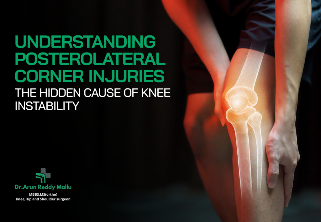
Understanding Posterolateral Corner Injuries – The Hidden Cause of Knee Instability When we think of knee injuries, the ACL (anterior cruciate ligament) and meniscus often take the spotlight. However, there’s a lesser-known yet crucial structure that plays a significant role in knee stability — the posterolateral corner (PLC). Injuries to this region are often overlooked or misdiagnosed, leading to chronic knee instability, pain, and difficulty returning to normal activities. In this article, we’ll explore what the posterolateral corner is, why injuries to it are so impactful, how they are diagnosed, and the modern treatment options available to restore knee stability. What Is the Posterolateral Corner (PLC)? The posterolateral corner of the knee is a complex network of ligaments, tendons, and muscles that stabilize the knee joint, especially against external rotation and backward movement of the tibia (shin bone). The key structures in the PLC include: Lateral collateral ligament (LCL):Provides stability on the outer side of the knee. Popliteus tendon:Helps control rotation of the knee and prevents excessive outward rolling. Popliteofibular ligament:Supports the popliteus tendon in preventing excessive motion. Arcuate ligament complex and posterolateral capsule:Additional supporting structures that add rotational and lateral stability. Together, these elements work in harmony with other ligaments like the ACL and PCL (posterior cruciate ligament) to maintain proper knee alignment and movement. How Do Posterolateral Corner Injuries Occur? PLC injuries usually happen due to high-energy trauma or sudden twisting forces on the knee. Common causes include: Sports injuries:Athletes in sports like football, soccer, basketball, and skiing are at higher risk. Sudden pivoting or a direct blow to the inside of the knee can stretch or tear the PLC structures. Motor vehicle accidents:A direct impact to the knee during a crash can lead to severe PLC injuries, often in combination withACL or PCL tears. Falls or hyperextension injuries:Landing awkwardly after a jump or an unexpected twist while running can overstretch the ligaments of the posterolateral corner. Combined ligament injuries:PLC injuries often don’t occur in isolation. They are commonly seen along with ACLor PCL tears, complicating both diagnosis and treatment. Symptoms of Posterolateral Corner Injuries Many people with PLC injuries may not realize the true source of their knee instability because symptoms often mimic those of other ligament injuries. Common signs include: Knee instability:A feeling that the knee “gives way” during walking or pivoting. Pain on the outer side of the knee:Especially noticeable when twisting or turning. Swelling and tenderness:Around the outer and back portion of the knee. Difficulty climbing stairs or changing direction:Due to loss of rotational control. Locking or clicking sensation:When the knee moves through its range of motion. If left untreated, these symptoms can worsen over time, leading to chronic instability and even early osteoarthritis. Why Are PLC Injuries Often Missed? PLC injuries can be challenging to diagnose because: The symptoms overlap with ACL or meniscus injuries. Standard imaging tests may not always clearly show damage to the smaller structures in the PLC. The focus is often on more commonly injured ligaments, leaving PLC tears unaddressed. When a PLC injury is missed, patients who undergo ACL reconstruction alone may continue to feel instability or have repeated graft failures. That’s why accurate diagnosis and comprehensive treatment are essential. Diagnosis of Posterolateral Corner Injuries Diagnosing PLC injuries requires a detailed clinical examination and advanced imaging. Clinical Examination An orthopedic specialist performs several tests to evaluate knee stability, including: Dial Test:Checks for excessive external rotation at the knee. Varus Stress Test:Assesses lateral stability of the knee joint. External Rotation Recurvatum Test:Evaluates both rotational and hyperextension instability. These tests help identify the specific ligaments and tendons affected. Imaging Tests MRI (Magnetic Resonance Imaging):The most accurate imaging tool to visualize soft tissue injuries. MRI can reveal tears in the LCL, popliteus tendon, or other PLC structures. X-rays:Used to detect any bone injuries or alignment issues. Stress Radiographs:May be used to assess the degree of joint laxity under stress. An experienced orthopedic surgeon will correlate clinical findings with imaging results to confirm the diagnosis and plan treatment. Grades of PLC Injury PLC injuries are classified into three grades based on severity: Grade I (Mild):Stretching of the ligaments with minimal instability. Grade II (Moderate):Partial tear with some joint laxity. Grade III (Severe):Complete tear of multiple structures leading to significant instability. Accurate grading is essential, as it determines whether conservative management or surgical reconstruction is needed. Treatment Options for Posterolateral Corner Injuries The treatment plan depends on the severity of the injury and whether other ligaments are also affected. Non-Surgical Treatment (For Mild Injuries) For Grade I or some Grade II injuries: Rest and immobilization:To allow the damaged tissues to heal. Physical therapy:Strengthening exercises for quadriceps, hamstrings, and hip muscles to improve knee stability. Bracing:A hinged knee brace may be recommended to restrict excessive movement. Gradual return to activity:Under the supervision of a physiotherapist. Non-surgical management can yield good results in minor injuries if rehabilitation is done correctly. Surgical Treatment (For Moderate to Severe Injuries) For Grade II (severe) and Grade III injuries, especially when combined with ACL or PCL tears, surgical reconstruction is often necessary. PLC Reconstruction: The surgeon reconstructs the damaged ligaments using grafts (from the patient’s own tissue or a donor). The goal is to restore the natural anatomy and function of the knee’s posterolateral corner. Surgery may be performed along with ACL or PCL reconstruction for complete joint stability. Post-Surgical Rehabilitation: Rehabilitation is a critical phase of recovery and typically includes: Controlled weight-bearing with crutches for the first few weeks. Range-of-motion exercises to prevent stiffness. Gradual strengthening and balance training. Return to sports or heavy physical activity usually after 6–9 months, depending on recovery. Early and precise surgical intervention significantly improves outcomes and helps prevent long-term instability. Potential Complications of Untreated PLC Injuries Ignoring or misdiagnosing a PLC injury can lead to: Persistent instability:Difficulty walking, running, or pivoting. Recurrent ligament tears:Especially if ACL reconstruction is done without addressing PLC damage. Joint degeneration:Leading to early onset osteoarthritis. Chronic pain and reduced performance:Particularly in athletes or active individuals. Hence, timely diagnosis and a multidisciplinary treatment approach are crucial for long-term knee health. Recovery
Is ACL Surgery Painful? What to Expect Before and After Surgery
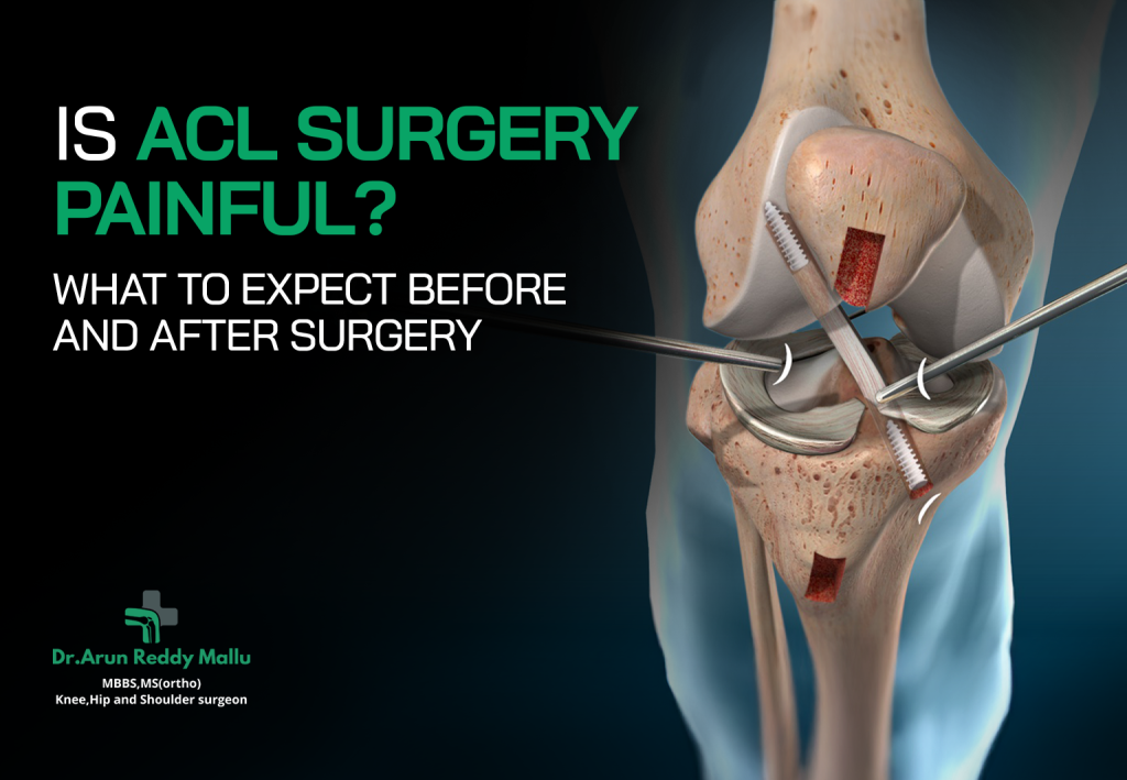
Is ACL Surgery Painful? What to Expect Before and After Surgery A torn Anterior Cruciate Ligament (ACL) is one of the most common knee injuries, especially among athletes and physically active individuals. If you’ve been advised to undergo ACL surgery, one of your biggest concerns is likely pain—both during and after the procedure. The good news is that modern surgical techniques and pain management strategies have made ACL reconstruction far less painful and more predictable than it used to be. In this detailed guide, we’ll walk you through what ACL surgery involves, how pain is managed, and what to expect before and after surgery for a smoother recovery journey. Understanding ACL and Its Role The ACL is one of the key ligaments in your knee joint that helps maintain stability during movement. It connects the thigh bone (femur) to the shin bone (tibia) and prevents the knee from sliding forward excessively. When the ACL tears—often due to sudden twisting motions, sports injuries, or accidents—it doesn’t heal on its own. In many cases, ACL reconstruction surgery becomes necessary to restore knee stability, prevent further damage, and allow a return to active living. Is ACL Surgery Painful? The Honest Answer During the Surgery:No, you won’t feel pain during the ACL surgery itself because it’s performed under spinal or general anesthesia. An anesthesiologist ensures you remain comfortable and pain-free throughout the procedure. After the Surgery:You can expect some discomfort, swelling, and soreness in the first few days following surgery. However, pain is effectively managed through medications, ice therapy, rest, and gradual rehabilitation exercises. With proper care and adherence to post-surgical guidelines, pain levels typically decrease significantly within the first two weeks, and most patients are able to walk with minimal discomfort soon after. What to Expect Before ACL Surgery Before undergoing ACL surgery, your orthopedic surgeon will guide you through several preparatory steps to ensure a successful outcome. Pre-Surgical Evaluation A complete assessment will be done, including: Physical examination MRI scan to confirm the ligament tear Discussion about your medical history and activity level Preoperative blood tests or health check-ups Prehabilitation (“Prehab”) Your doctor may recommend physiotherapy before surgery. Strengthening your quadriceps, hamstrings, and core muscles beforehand helps speed up recovery later. This “prehab” phase also reduces stiffness and improves flexibility. Surgery Day Preparation On the day of surgery: You’ll be asked to fast for several hours. The anesthesiologist will explain the type of anesthesia used. You’ll be shifted to the operating room, where the surgery typically takes about 5 to 2 hours. The Procedure ACL reconstruction involves replacing the torn ligament with a tissue graft (usually taken from your own hamstring or patellar tendon). The graft acts as a scaffold for new ligament growth over time. The surgery is done arthroscopically — through small incisions using a camera and tiny instruments, minimizing pain and scarring. What to Expect After ACL Surgery Recovery after ACL surgery is a gradual process, and understanding each stage helps set realistic expectations. Immediately After Surgery You’ll spend a few hours in the recovery room as anesthesia wears off. Your knee will likely be bandaged, and a brace may be used to keep it stable. Pain medication will be given to manage initial discomfort. The First Few Days Pain and Swelling:It’s normal to experience mild to moderate pain and swelling. Applying ice packs and keeping your leg elevated can help reduce this. Mobility:Crutches are used to avoid putting weight on your operated leg initially. Medications:Your doctor will prescribe antibiotics and painkillers to prevent infection and ease discomfort. The First Two Weeks During this period, the focus is on managing pain, reducing swelling, and regaining knee motion. Physiotherapists guide you through gentle exercises like ankle pumps and leg raises to maintain circulation and muscle tone. You may still feel soreness, especially when bending or straightening your leg, but this is part of the healing process. Following your doctor’s instructions closely will make a big difference in comfort and recovery speed. Pain Management After ACL Surgery Pain levels vary from person to person, but with modern approaches, most patients describe their pain as manageable rather than severe. Common methods include: Medications Pain-relieving medicines like NSAIDs, acetaminophen, or prescribed opioids (for short-term use) help control discomfort. Your doctor will adjust doses to balance relief and safety. Cold Therapy Applying ice or using a cold compression device reduces inflammation and swelling significantly in the first few days. Elevation and Rest Keeping the leg elevated promotes blood circulation and minimizes pressure around the knee joint, leading to less pain and stiffness. Physiotherapy Regular, guided physiotherapy sessions not only improve strength and flexibility but also prevent pain caused by stiffness or muscle weakness. Nerve Blocks (Optional) Some patients receive a nerve block injection around the knee before or after surgery. This numbs the area for several hours, providing additional pain control immediately after surgery. Physical Therapy and Recovery Timeline Rehabilitation is the most crucial part of ACL recovery. Here’s a general timeline: Weeks 1–2:Focus on gentle mobility, swelling reduction, and light exercises. Weeks 3–6:You’ll start weight-bearing activities and work on strengthening the quadriceps and hamstrings. Months 2–4:More active physiotherapy — including balance, endurance, and controlled movements. Months 5–6:Gradual return to jogging, running, or light sports under supervision. After 6 Months:Most patients return to normal activities, though full sports performance may take 8–9 months depending on individual progress. Throughout the process, communication with your physiotherapist and surgeon ensures proper healing and prevents complications. Common Concerns After ACL Surgery How long will I need painkillers?Most patients use prescription pain medication for about a week, then switch to milder pain relievers as needed. When can I walk without crutches?Typically within 2–3 weeks, depending on your strength and stability. Is there pain during physiotherapy?Some mild discomfort is expected, but it should not be severe. Your therapist adjusts exercises to your tolerance level. Can ACL surgery fail?Though rare, improper rehabilitation or premature return to sports can lead to graft failure. Following medical advice is key to preventing this. Tips for a Smooth Recovery Follow all
Shoulder Arthroscopy – Procedure, Benefits & Conditions Treated
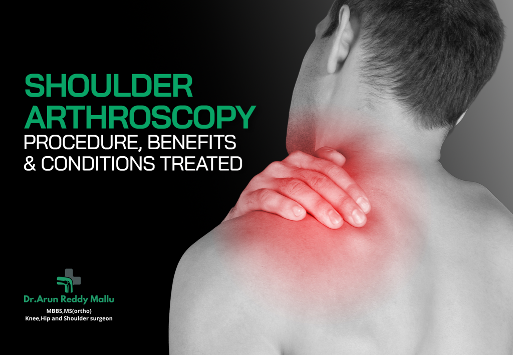
Shoulder Arthroscopy – Procedure, Benefits & Conditions Treated Shoulder pain, stiffness, or restricted motion can severely affect your quality of life, especially if you’re active or perform repetitive arm movements. Over time, injuries, wear and tear, or degenerative changes can lead to shoulder problems that require medical attention. Fortunately, advances in orthopedic surgery have made it possible to diagnose and treat many of these conditions using a minimally invasive technique known as shoulder arthroscopy. What Is Shoulder Arthroscopy? Shoulder arthroscopy is a surgical procedure that allows orthopedic surgeons to view and treat the inside of the shoulder joint using a small camera called an arthroscope. Unlike traditional open surgery that requires large incisions, arthroscopy is performed through tiny keyhole-sized cuts. The arthroscope projects real-time images onto a screen, helping the surgeon see the joint structures clearly and perform repairs using miniature surgical instruments. Because it causes less trauma to tissues, shoulder arthroscopy often leads to faster healing, smaller scars, and reduced pain compared to open surgery. It’s now one of the most common and effective procedures used to treat a wide range of shoulder conditions. Why Is Shoulder Arthroscopy Done? The shoulder is the most mobile joint in the body. It’s formed by the humeral head (ball) fitting into the glenoid cavity (socket) of the shoulder blade. This mobility makes it vulnerable to injury and instability. Overuse, accidents, or degeneration can damage the joint’s soft tissues — such as the rotator cuff, labrum, ligaments, tendons, and cartilage — causing pain, weakness, or restricted movement. When conservative treatments like medication, physiotherapy, or injections fail to relieve symptoms, arthroscopy is recommended. It allows the surgeon to both diagnose and correct the problem in the same sitting, ensuring long-term relief and restoration of shoulder function. Common Shoulder Conditions Treated with Arthroscopy Rotator Cuff Tears Rotator cuff injuries are among the most frequent reasons for shoulder arthroscopy. The rotator cuff consists of four tendons that stabilize and move the shoulder joint. Tears may result from age-related degeneration or trauma.Arthroscopic rotator cuff repair involves reattaching the torn tendon to the bone using small anchors and sutures. It restores shoulder strength, reduces pain, and improves range of motion. Shoulder Impingement and Bursitis In shoulder impingement, soft tissues like tendons or the bursa become trapped under the shoulder blade, leading to inflammation and pain during movement. Arthroscopy can smooth bone edges and remove inflamed tissue (a process known as subacromial decompression) to relieve impingement. Labral Tears and Shoulder Instability The labrum is a ring of cartilage that deepens the shoulder socket and keeps the joint stable. Injuries or dislocations can tear the labrum, causing instability, clicking, or recurrent shoulder dislocations. Arthroscopic labral repair reattaches the torn cartilage using sutures, restoring stability. Frozen Shoulder (Adhesive Capsulitis) Frozen shoulder causes pain and stiffness due to thickening and tightening of the joint capsule. When non-surgical treatments fail, arthroscopic capsular release can loosen the tight tissues and restore mobility. Loose Bodies or Cartilage Damage Small fragments of cartilage or bone may float inside the shoulder joint, causing locking or catching sensations. Arthroscopy allows for precise removal of these loose bodies and smoothing of rough cartilage. Biceps Tendon Disorders Tears or inflammation of the biceps tendon can cause pain at the front of the shoulder. Arthroscopic procedures such as biceps tenodesis (reattaching the tendon) or tenotomy (releasing it) help alleviate symptoms. Shoulder Arthritis (Early Stages) While advanced arthritis may require replacement surgery, early arthritis can be managed arthroscopically by cleaning the joint (debridement) and removing inflamed tissue to reduce discomfort. Step-by-Step Overview of the Shoulder Arthroscopy Procedure Pre-Surgical Preparation Before the procedure, the patient undergoes: Detailed physical examination X-rays and MRI scans to assess the shoulder Medical evaluation for anesthesia fitness Patients are advised to fast before surgery and stop certain medications (like blood thinners) as per doctor’s advice. Anesthesia and Positioning The surgery is typically performed under regional or general anesthesia. The patient may be placed in either the beach-chair or side-lying position, depending on the surgeon’s preference. Creating Portals Small incisions (portals) are made around the shoulder. Through one incision, the arthroscope is inserted, and through others, surgical instruments are passed. Sterile fluid is used to expand the joint for better visualization. Diagnostic Examination The surgeon first performs a diagnostic inspection to identify all internal damage — examining the rotator cuff, labrum, cartilage, biceps tendon, and capsule. Performing Repairs Based on findings, various procedures may be carried out: Rotator cuff repairusing suture anchors Labral repairfor instability Subacromial decompressionto remove bone spurs Capsular releasefor frozen shoulder Debridementor tenodesis for biceps injuries Once repairs are completed, the instruments are withdrawn, and the small incisions are closed with sutures or adhesive strips. Recovery and Rehabilitation Patients are usually discharged on the same day or the next morning. A sling is worn for support during the initial healing phase. Pain and swelling are managed with medication and ice packs. Physiotherapybegins within a few days to restore motion and strength. Recovery time depends on the extent of repair — simple debridements may heal in a few weeks, while complex repairs may take 3–6 months for full recovery. Benefits of Shoulder Arthroscopy Minimally Invasive– Smaller incisions mean less tissue damage and scarring. Reduced Pain and Swelling– Patients experience significantly less postoperative discomfort. Faster Recovery– Early mobilization and quicker return to daily activities. Lower Infection Risk– Smaller wounds reduce the risk of infection or complications. Improved Precision– The arthroscope provides a magnified view, allowing highly accurate repairs. Same-Day Discharge– Most procedures are performed as outpatient surgeries. Better Cosmetic Results– Tiny scars are barely noticeable. These advantages make arthroscopy a preferred choice for both surgeons and patients. Possible Risks and Complications Though considered safe, shoulder arthroscopy carries some minor risks, such as: Temporary stiffness or restricted motion Bleeding or fluid accumulation Infection (rare) Nerve irritation or numbness (usually temporary) Re-tear or incomplete healing (in large or chronic injuries) Following the surgeon’s rehabilitation protocol strictly can significantly minimize these risks and ensure optimal recovery. Who Is an Ideal Candidate? You may be a
A Comprehensive Guide to AVN Hip and Its Modern Treatment Options
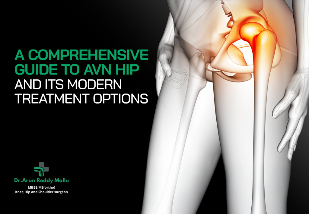
A Comprehensive Guide to AVN Hip and Its Modern Treatment Options Avascular Necrosis (AVN) of the hip, also known as osteonecrosis, is a serious condition that occurs when blood supply to the hip bone (femoral head) is reduced or cut off. Without adequate blood flow, the bone tissue begins to die, leading to pain, limited mobility, and eventually, collapse of the joint surface. Early diagnosis and modern treatment approaches can significantly improve outcomes and preserve joint function. In this guide, we’ll explore what AVN hip is, its causes, symptoms, stages, and the most advanced treatment options available today. We’ll also highlight how Dr. Arun Reddy Mallu, an expert orthopaedic surgeon at Continental Hospitals, Hyderabad, is helping patients regain mobility and lead pain-free lives. What is AVN of the Hip? The hip joint is a ball-and-socket joint where the femoral head (ball) fits into the acetabulum (socket). In Avascular Necrosis (AVN), the blood flow to the femoral head is disrupted. Over time, the bone tissue deteriorates, leading to structural collapse of the joint and painful arthritis. The condition often affects adults between 30 and 50 years of age and can occur in one or both hips. Common Causes of AVN Hip Several factors can lead to AVN of the hip, including: Trauma or Injury:A fracture or dislocation of the hip can damage nearby blood vessels, restricting blood flow to the femoral head. Long-Term Steroid Use:Prolonged use of corticosteroids (like prednisone) can cause fat deposits in blood vessels, reducing blood circulation to bones. Alcohol Abuse:Excessive alcohol intake can increase lipid levels, leading to blockages in blood vessels supplying the bone. Medical Conditions:Certain health conditions, such as lupus, sickle cell anemia, diabetes, and pancreatitis, are associated with AVN. Idiopathic (Unknown Causes):In many cases, AVN develops without a clear cause, highlighting the importance of early screening and diagnosis. Early Symptoms of AVN Hip Recognizing early symptoms is crucial for preventing joint collapse and long-term disability. Common signs include: Persistent or intermittent groin painthat may extend to the thigh or buttock Stiffness and reduced range of motionin the hip Pain during walking or weight-bearing activities Limpingdue to pain or weakness Discomfort even at rest or while lying down in advanced stages When such symptoms appear, timely consultation with a specialist Dr. Arun Reddy Mallu at Continental Hospitals can make a significant difference in treatment outcomes. Stages of AVN Hip AVN progresses through distinct stages, from mild to severe: Stage I – Early Stage:No visible damage on X-rays, but MRI may show changes in blood supply. Stage II – Intermediate Stage:Bone structure starts to weaken; pain intensifies during activity. Stage III – Advanced Stage:Femoral head begins to collapse, leading to joint deformity. Stage IV – Late Stage:Severe arthritis develops, causing constant pain and limited movement. Early intervention during the first two stages offers the best chance of preserving the natural hip joint. Diagnosis of AVN Hip Accurate diagnosis is key to choosing the right treatment. A detailed medical history, physical examination, and imaging tests help confirm AVN. Common diagnostic tools include: X-rays:Useful for detecting bone collapse in later stages. MRI (Magnetic Resonance Imaging):The most sensitive test for early detection of AVN. CT Scan:Provides a detailed view of bone structure and joint damage. Bone Scans:Show reduced bone activity or poor blood flow in affected areas. At Continental Hospitals, specialists Dr. Arun Reddy Mallu use advanced imaging and diagnostic tools to determine the exact stage and extent of AVN for a personalized treatment plan. Modern Treatment Options for AVN Hip Treatment for AVN hip focuses on stopping disease progression, relieving pain, and restoring mobility. Depending on the stage and severity, the following treatment approaches are used: Non-Surgical Treatments (Early Stages) In early AVN cases, non-surgical management can help slow progression and preserve bone structure. Medications: Bisphosphonates to slow bone damage Anti-inflammatory drugs (NSAIDs) for pain relief Vasodilators or anticoagulants to improve blood flow Lifestyle Modifications: Reducing weight-bearing activities Using walking aids to reduce hip pressure Limiting alcohol and steroid use Physical Therapy: Customized exercises can help maintain joint function and strengthen surrounding muscles. Core Decompression Surgery This is a minimally invasive surgical option where small channels are drilled into the femoral head to reduce pressure and promote new blood vessel formation.Core decompression works best in early AVN stages before bone collapse occurs. Bone Grafting Bone grafts are used to replace dead bone tissue and stimulate regeneration. There are two types: Autograft:Bone taken from the patient’s own body Allograft:Bone from a donor This method is often combined with core decompression for better results. Stem Cell Therapy and Regenerative Medicine Modern medicine now uses stem cell therapy to regenerate damaged bone tissue and restore blood supply. Stem cells derived from bone marrow are injected into the femoral head to promote healing. This innovative technique offers promising results for early-stage AVN and is being successfully practiced by specialists in the field. Total Hip Replacement (THR) In advanced cases where the femoral head has collapsed, Total Hip Replacement surgery in Hyderabad is considered the gold standard. The damaged bone and cartilage are replaced with artificial components, allowing smooth and pain-free movement. Modern implants and robotic-assisted technology in Hyderabad ensure precise alignment, faster recovery, and longer implant lifespan. Recovery and Rehabilitation Recovery depends on the severity of AVN and the chosen treatment. After non-surgical treatments, patients can usually return to normal activities within a few weeks. Following core decompression or stem cell therapy, full recovery may take 3–6 months. For total hip replacement, rehabilitation may take up to 6 months with physical therapy. A structured rehab plan and regular follow-ups with your orthopaedic specialist are vital for long-term success. Preventing AVN Hip While not all causes can be avoided, some preventive measures include: Limiting alcohol and tobacco consumption Using steroids only under medical supervision Managing chronic health conditions such as diabetes or lupus Staying active and maintaining a healthy weight Regular orthopaedic check-ups can help detect early changes in bone health and prevent complications. Why Choose Dr. Arun Reddy Mallu at Continental Hospitals? Dr. Arun Reddy Mallu,
Bilateral Knee Replacement: Is There a Higher Risk of Complications?
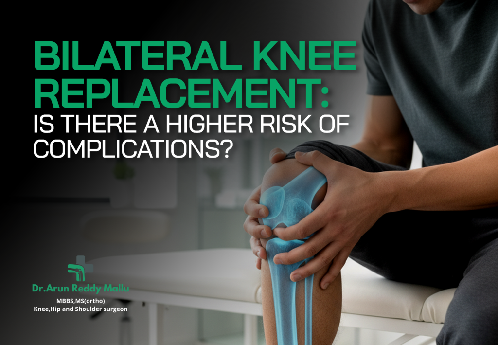
Bilateral Knee Replacement: Is There a Higher Risk of Complications? Bilateral knee replacement — replacing both knees during the same surgery — can be life-changing for people with severe arthritis in both knees. The thought of getting rid of pain in both joints at once is appealing: one hospital stay, one anesthesia, and recovery for both knees together. However, an important question arises: is there a higher risk of complications compared with operating on one knee at a time? This article explains the risks, who is a good candidate, how surgeons minimize complications, and what recovery involves. It also provides guidance on when to consider bilateral knee replacement surgery in Hyderabad. What is Bilateral Knee Replacement? Bilateral knee replacement means replacing both knee joints, either during the same surgical session (simultaneous) or through two separate operations (staged). Simultaneous bilateral replacement: Both knees are replaced at the same time under a single anesthesia. Staged bilateral replacement: One knee is replaced first, followed by the other after weeks or months of recovery. The ultimate goal in both approaches is the same: to relieve pain, restore function, and improve mobility. The key difference lies in timing and recovery demands. Is There a Higher Risk of Complications? Yes, simultaneous bilateral knee replacement is generally associated with a higher risk of complications compared with a single knee replacement. The reason is simple — the surgery is longer, involves both knees, and places greater physiological stress on the body. However, this does not mean it is unsafe. For the right patient, performed in an experienced center, outcomes can be excellent. Some of the more significant complications include: Cardiopulmonary events: Longer surgery and anesthesia can increase the risk of heart problems (like arrhythmia or heart attack) or breathing difficulties, particularly in patients with pre-existing conditions. Blood loss and transfusion: Operating on both knees at once can cause more blood loss, which may require a transfusion. Deep vein thrombosis (DVT) and pulmonary embolism (PE): The risk of blood clots is higher after major joint surgery. Operating on both knees raises that exposure further. Infection or wound issues: Because both knees are operated on simultaneously, there is a slightly higher chance of wound complications or infection. Postoperative confusion or complications in older adults: Longer anesthesia and hospital stays may increase risks like delirium or other medical problems in frail patients. Rehabilitation challenges: Both knees healing at once means more demanding rehabilitation, requiring stronger support systems at home. It is important to note that with modern surgical techniques, excellent anesthesia, and better recovery protocols, these risks are greatly reduced when the surgery is done in high-quality hospitals. Who is a Good Candidate? Not every patient with arthritis in both knees is an ideal candidate for simultaneous bilateral replacement. Doctors carefully evaluate medical history and fitness before recommending it. Generally, it may be considered for patients who: Are otherwise healthy and physically strong. Have no major uncontrolled heart, lung, or kidney problems. Are younger or middle-aged with good overall fitness. Have strong support at home for rehabilitation. Wish to complete treatment in one hospital stay rather than returning for a second surgery. Patients who are older, frail, or have serious health conditions may be advised to undergo staged knee replacement instead, reducing stress on the body. How Surgeons Reduce Risks Modern medicine offers several ways to minimize risks and improve recovery outcomes in bilateral knee replacement: Thorough Preoperative Assessment: Patients undergo detailed evaluations, including cardiac and pulmonary testing, to ensure fitness for surgery. Medical Optimization: Pre-existing conditions such as diabetes, high blood pressure, or anemia are carefully managed before the operation. Blood Conservation Techniques: Methods like tranexamic acid help reduce blood loss. Preventing Blood Clots: Both mechanical (compression devices) and medication-based methods are used. Enhanced Recovery Protocols: Early mobilization, multimodal pain relief, and structured physiotherapy are key to safe recovery. Experienced Surgical and Anesthesia Teams: Choosing a high-volume center with specialists experienced in bilateral procedures significantly reduces complication risks. Recovery After Bilateral Knee Replacement Recovery after bilateral knee replacement requires dedication and support. Patients usually start walking with assistance within one to two days after surgery. The hospital stay may be slightly longer than for single knee replacement, depending on progress. Physiotherapy begins earlyto help restore mobility. Pain control protocolsallow patients to exercise and move without severe discomfort. The first 6–12 weeksare the most crucial for regaining strength and independence. Long-term recoverymay take 6–12 months, after which most patients enjoy significant pain relief and improved quality of life. Support at home is critical, as patients are managing both knees at once. Family or caregiver assistance during the early recovery period is highly beneficial. Key Considerations Before Surgery If you are considering bilateral knee replacement, here are some important steps to take: Comprehensive medical evaluation— check your heart, lungs, and other organ systems. Discuss with your knee specialistwhether simultaneous surgery is suitable for your condition. Ask about the hospital’s experiencewith bilateral knee replacements. Prepare your home environment— ensure it is safe and supportive for recovery. Follow prehabilitation exercisesrecommended by your physiotherapist to strengthen muscles before surgery. Expert Knee Care in Hyderabad For patients exploring bilateral knee replacement Surgery in Hyderabad, choosing the right surgeon and hospital makes all the difference. Dr. Arun Reddy Mallu, an experienced knee specialist doctor in Hyderabad, provides expert care and guidance at Continental Hospitals. With his expertise in advanced knee replacement procedures, patients receive individualized treatment plans that prioritize safety, effective pain relief, and long-term function. Conclusion Bilateral knee replacement does carry a higher risk of complications compared with operating on one knee at a time. The longer surgery, greater blood loss, and rehabilitation demands mean that only carefully selected patients are suitable for this approach. However, in the right hands and with proper medical preparation, it can be safe and rewarding, helping patients regain mobility and independence more efficiently. If you are considering this procedure, consult with an experienced specialist like Dr. Arun Reddy Mallu at Continental Hospitals, Hyderabad. With expert guidance and modern medical
Does Running Damage Your Knees or Strengthen Them?
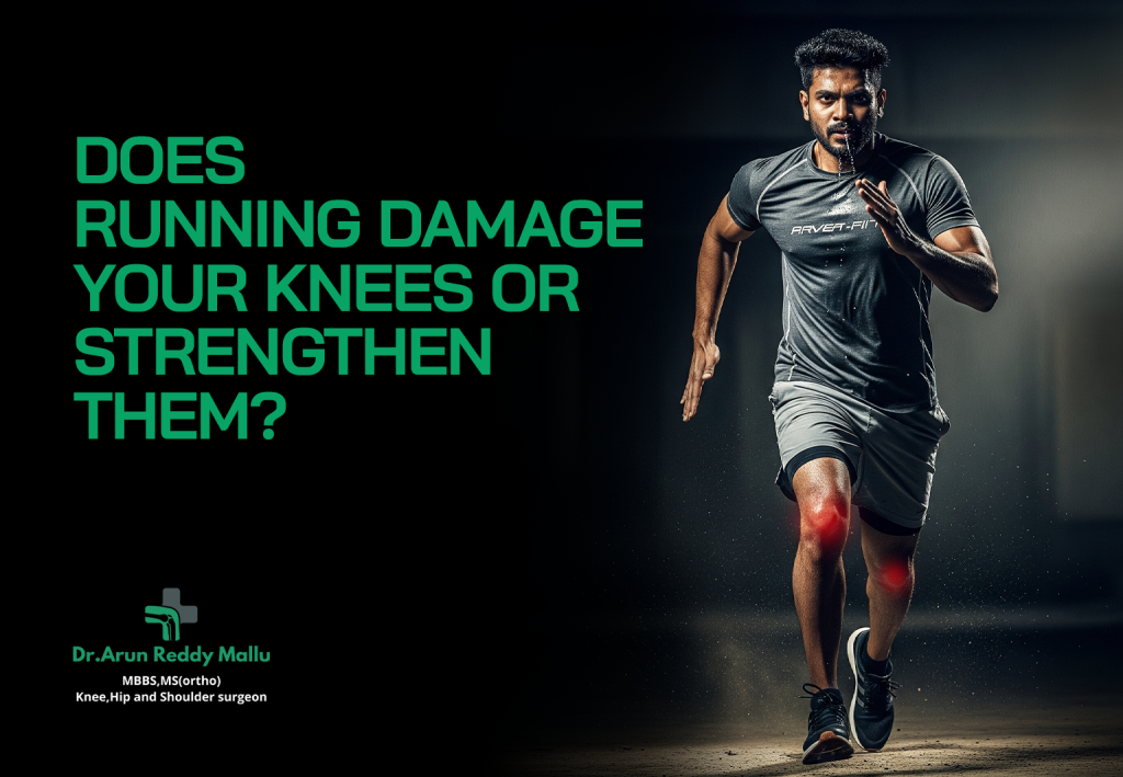
Does Running Damage Your Knees or Strengthen Them? Running is one of the most popular forms of exercise across the world. From early morning joggers to seasoned marathoners, millions rely on running as a way to maintain their fitness, manage weight, and improve cardiovascular health. Yet, one question has lingered for decades: does running damage your knees, or does it strengthen them? Many people avoid running because of the fear that it will “wear out” their knee joints and eventually lead to arthritis. On the other hand, countless runners swear by the benefits, claiming that running has kept their knees strong and their bodies active for years. So, what does science say? Let’s explore this fascinating debate. Understanding the Knee Joint Before diving into the effects of running, it’s important to understand how the knee works.The knee joint is the largest joint in the human body. It is a hinge joint, connecting the thigh bone (femur) to the shin bone (tibia), with the kneecap (patella) protecting it in front. Supporting structures like ligaments, tendons, and cartilage help stabilize the joint and absorb shock during movements like walking, running, or jumping. If you ever face issues related to these structures, consulting a knee specialist in Hyderabad can help with accurate diagnosis and treatment. Since running involves repetitive impact and load-bearing, the knee joint plays a critical role in absorbing forces that are several times your body weight with each stride. This is why concerns about knee damage often arise. The Common Myth: Running Causes Arthritis The most widespread belief is that running “wears out” the knee cartilage, causing osteoarthritis—a condition where the protective cartilage gradually breaks down, leading to pain, stiffness, and reduced mobility. However, numerous studies over the past two decades have challenged this assumption. Research has shown that recreational running does not increase the risk of knee arthritis. In fact, some studies suggest that moderate running may even reduce the risk compared to sedentary individuals. The key lies in differentiating between recreational runners, who typically run moderate distances regularly, and elite or professional athletes, who may be at risk due to extremely high mileage and repetitive overuse. How Running Can Strengthen Your Knees Strengthening Muscles Around the Knee Running activates and strengthens key muscle groups, including the quadriceps, hamstrings, calves, and hip muscles. Stronger muscles provide better support and stability to the knee joint, reducing the risk of injuries. Improved Bone Density Weight-bearing activities like running help increase bone density. This makes your knee bones more resilient and less likely to suffer stress fractures or weakness over time. Cartilage Health Contrary to popular belief, running can stimulate cartilage by improving nutrient delivery. Cartilage does not have a direct blood supply; instead, it receives nutrients through the movement of synovial fluid during joint activity. Regular running encourages this process, keeping the cartilage healthier. Weight Management One of the biggest risk factors for knee arthritis is excess body weight. Running helps burn calories and maintain a healthy weight, thereby reducing unnecessary strain on the knees. When Running Can Harm Your Knees While recreational running has many benefits, it’s not without risks. Poor technique, inadequate footwear, or excessive mileage can lead to knee problems. Some common running-related knee issues include: Runner’s Knee (Patellofemoral Pain Syndrome) This condition causes pain around the kneecap, especially when running downhill, climbing stairs, or sitting for long periods. It’s often due to overuse, muscle imbalances, or improper running mechanics. Iliotibial Band Syndrome (ITBS) The iliotibial band is a thick band of tissue running along the outside of the thigh. When it becomes tight, it can rub against the outer knee, causing pain. Meniscus Injuries Sudden twisting or impact during running can sometimes tear the meniscus (cartilage in the knee). Though not always linked to running alone, it’s a risk when combined with poor form or uneven surfaces. Overuse Injuries High-mileage training without adequate rest increases the risk of micro-injuries in the knee and surrounding tissues. Factors That Influence Whether Running Helps or Hurts Your Knees Running Technique Proper form—like landing softly on your midfoot instead of striking heavily on your heels—can reduce knee stress. Maintaining good posture and cadence also matters. Footwear Worn-out or improper running shoes can alter your gait and increase knee strain. Investing in supportive shoes tailored to your foot type makes a big difference. Running Surface Hard surfaces like concrete create higher impact forces compared to softer tracks or trails. Mixing up surfaces can reduce repetitive stress. Training Volume and Intensity Sudden increases in distance or speed can overload the knees. Gradual progression allows your joints and muscles to adapt safely. Strength and Flexibility Runners who neglect strength training and stretching are more prone to knee pain. Strengthening the hips, core, and legs improves knee stability. What the Research Says Several large studies have analyzed the long-term impact of running on knee health: A 2017 study published in Arthritis Care & Researchfound that recreational runners had lower rates of knee osteoarthritis compared to non-runners. Another study in the Journal of Orthopaedic & Sports Physical Therapyreported that running may reduce inflammation in the knee joint, contrary to the assumption that it worsens it. Long-term runners who maintain moderate mileage tend to have healthier knees than those who are sedentary, likely due to the protective effects of muscle strength, cartilage stimulation, and weight management. These findings suggest that moderate running is not only safe for the knees but may actually protect them in the long run. Tips for Protecting Your Knees While Running If you enjoy running but are worried about your knees, here are some practical tips: Warm Up and Cool Down– Gentle stretching and light jogging before and after runs can reduce injury risk. Strength Train– Incorporate exercises like squats, lunges, and leg presses to build supportive muscles. Invest in Good Shoes– Replace them every 500–800 km, or when cushioning wears out. Run on Varied Surfaces– Alternate between grass, trails, and treadmill to reduce repetitive stress. Listen to Your Body– If you experience
Partial Knee Replacement Options: Fixed vs. Mobile Bearing Explained
Partial Knee Replacement Options: Fixed vs. Mobile Bearing Explained When knee pain from arthritis or injury begins to interfere with daily life, many people assume a total knee replacement is the only solution. However, not every patient requires complete replacement of the knee joint. For those with damage limited to a single compartment of the knee, Partial Knee Replacement (PKR) can be a highly effective treatment option. Within partial knee replacement, two major implant designs are widely used: Fixed Bearing and Mobile Bearing implants. Both options have distinct features, benefits, and limitations. Choosing between them depends on factors such as age, activity level, bone structure, and the extent of knee damage. What is Partial Knee Replacement? The knee joint has three compartments: Medial (inner side of the knee) Lateral (outer side of the knee) Patellofemoral (between the kneecap and thigh bone) When arthritis or cartilage damage affects only one compartment, surgeons may recommend partial knee replacement instead of a total knee replacement. In this surgery: Only the damaged portion of the knee is replaced with an artificial implant. Healthy bone, cartilage, and ligaments in the other compartments are preserved. Patients typically recover faster with less pain and improved joint function. This procedure allows for a more natural feel in the knee compared to total knee replacement, as much of the original joint structure remains intact. Fixed Bearing vs. Mobile Bearing: An Overview The main difference between fixed bearing and mobile bearing partial knee replacement lies in how the artificial components interact and move. Fixed Bearing Implant In a fixed bearing design, the plastic insert (polyethylene) is attached firmly to the metal tibial (shin bone) component. The femoral (thigh bone) component glides directly over this fixed insert. Movement occurs between the femoral component and the polyethylene, not within the insert itself. Advantages: Simpler design with a proven track record. Lower risk of dislocation since the insert doesn’t move. Generally less expensive compared to mobile bearing implants. Suitable for older or less active patients who don’t place high demands on the joint. Limitations: Higher risk of wear and tear in the polyethylene insert, especially in younger and more active patients. May place more stress on surrounding bone and tissue, leading to loosening over time. Mobile Bearing Implant In this design, the polyethylene insert is not fixed but can rotate or move slightly on the tibial baseplate. The femoral component glides over this mobile insert, allowing more natural joint movement. Advantages: Mimics the natural rolling and gliding motion of the knee more closely. Distributes stress more evenly, reducing wear on the polyethylene. Longer-lasting in younger, active patients compared to fixed bearing. Provides better range of motion and potentially a more natural-feeling knee. Limitations: Slightly more complex surgical technique required. Risk of dislocation of the insert if not placed precisely. Typically more expensive than fixed bearing options. Who is the Right Candidate? Choosing between fixed and mobile bearing implants depends on several factors: Age and Activity Level Fixed bearing: Best suited for patients over 65 or those with lower activity levels. Mobile bearing: Ideal for younger, more active patients who want better motion and longevity. Bone Quality Mobile bearing implants require strong, stable bone support. Fixed bearing implants can tolerate slightly weaker bone. Weight Considerations Heavier patients may benefit from mobile bearing implants due to their ability to reduce wear. However, implant dislocation risk must be carefully considered. Surgeon’s Expertise Since mobile bearing implants require precise alignment, outcomes often depend on the surgeon’s skill and experience. Advantages of Partial Knee Replacement Over Total Replacement Regardless of implant type, partial knee replacement has several benefits compared to total knee replacement: Less invasive: Smaller incisions and less tissue disruption. Faster recovery: Many patients resume normal activities within weeks. Natural knee function: Preserves ligaments and unaffected compartments. Reduced pain and stiffness: Most patients report significant improvement. Longevity: Modern implants last 10–20 years, depending on patient lifestyle. Risks and Considerations Like any surgery, partial knee replacement carries some risks, such as: Infection Blood clots Implant loosening or wear Continued progression of arthritis in other compartments Patients should carefully discuss risks, benefits, and implant options with their orthopedic surgeon before making a decision. Conclusion Both Fixed Bearing and Mobile Bearing implants in partial knee replacement offer excellent results, but the right choice depends on your age, activity level, bone health, and the surgeon’s recommendation. If you’re older, less active, or prefer a simpler, cost-effective solution, Fixed Bearingimplants may be ideal. If you’re younger, more active, and want a natural-feeling knee with long-term durability, Mobile Bearingimplants could be the better option. Discover Partial Knee Replacement options with Dr. Arun Reddy Mallu at Continental Hospital — call +91 99591 35963 for expert consultation.
Will Your ACL Heal on Its Own or Do You Need Surgery?

Will Your ACL Heal on Its Own or Do You Need Surgery? The anterior cruciate ligament (ACL) is one of the most important ligaments in the knee joint. It plays a vital role in stabilizing the knee, allowing us to run, jump, pivot, and perform activities that require quick changes in direction. Unfortunately, ACL injuries are among the most common sports-related injuries worldwide, often occurring in athletes, fitness enthusiasts, and even in people performing daily activities. One of the biggest concerns after an ACL tear is whether the ligament can heal on its own or whether surgical intervention is necessary. The answer isn’t always straightforward—it depends on the severity of the injury, your activity level, and your personal goals for mobility. Understanding the ACL and Its Role The ACL is a strong band of tissue that connects the thighbone (femur) to the shinbone (tibia). It prevents excessive forward movement of the tibia relative to the femur and provides stability when you twist or pivot. When the ACL tears, either partially or completely, the knee loses this stability. Patients often report hearing a “pop” sound at the time of injury, followed by pain, swelling, and difficulty bearing weight. Can the ACL Heal on Its Own? Unlike some ligaments in the body, the ACL has limited healing ability. This is because of its poor blood supply. While minor sprains or partial tears may improve with rest, physical therapy, and rehabilitation, a complete ACL tear rarely heals on its own. Factors that influence healing include: Type of tear– A small or partial tear may respond to non-surgical treatment, while complete tears usually require surgery. Age– Younger individuals with high physical demands may require surgical repair to prevent future instability. Activity level– Athletes or those engaged in heavy physical activity often need surgery for a stable and functional knee. Associated injuries– ACL injuries are often accompanied by damage to meniscus or cartilage, which may require surgical intervention. Symptoms of an ACL Injury Recognizing the signs of an ACL tear is critical to seeking timely treatment. Common symptoms include: A popping sensation at the time of injury Rapid swelling within hours Severe knee pain Instability or “giving way” feeling while walking or pivoting Limited range of motion If these symptoms are present, it’s important to consult an orthopedic specialist as soon as possible. Non-Surgical Management of ACL Injuries Not all ACL injuries require surgery. In some cases, conservative management can be effective, especially for patients who do not engage in high-intensity sports. Non-surgical options include: Rest and activity modification– Avoiding movements that stress the knee. Physical therapy– Strengthening surrounding muscles (quadriceps, hamstrings) to provide better knee support. Bracing– Knee braces may help provide stability during movement. Medication– Anti-inflammatory drugs for pain and swelling. This approach may be suitable for: Patients with partial tears Older adults with lower activity demands Individuals not involved in pivot-heavy sports However, one must note that without surgery, the risk of long-term instability and further damage to the knee joint remains. When is Surgery Needed? Surgery is usually recommended when: The ACL is completely torn– Without reconstruction, the knee often remains unstable. Patients are young and active– Athletes or those who wish to continue sports usually require surgical repair. Associated injuries are present– Torn meniscus or cartilage damage often need correction during ACL surgery. Daily function is affected– If the knee frequently gives way during walking or simple tasks. ACL reconstruction surgery involves replacing the torn ligament with a graft, often taken from the patient’s hamstring or patellar tendon. With proper rehabilitation, most patients can return to sports and active lifestyles. Recovery Timeline After ACL Surgery Recovery from ACL reconstruction requires patience and discipline. On average: First 2 weeks:Focus on reducing swelling, regaining basic motion. Weeks 3–6:Begin strengthening exercises, start walking with minimal assistance. Months 3–6:Progressive strengthening, running, agility training. Month 9–12:Return to sports and full activity after clearance from the surgeon. Adhering to a structured rehabilitation program is as important as the surgery itself for long-term success. Risks of Avoiding Surgery For patients with complete ACL tears who opt out of surgery, the long-term risks include: Chronic instability Higher chance of re-injury Increased wear and tear on the meniscus and cartilage Greater risk of developing osteoarthritis at an earlier age This is why it is important to consult a qualified orthopedic surgeon who can assess your case and guide you on the right treatment path. Role of an Orthopedic Specialist Since ACL injuries can have lasting consequences, consulting an experienced orthopedic surgeon is crucial. A specialist can perform diagnostic tests such as MRI and physical examinations (like the Lachman test) to determine the extent of damage. One such expert is Dr. Arun Reddy Mallu at Continental Hospital, a well-known orthopedic surgeon with extensive experience in treating ACL injuries and other knee-related conditions. His expertise in advanced surgical techniques and patient-focused rehabilitation ensures that individuals can regain mobility and return to their normal lifestyles with confidence. Preventing ACL Injuries While not all ACL injuries can be prevented, strengthening the knee and practicing safe movement can reduce risks. Some preventive strategies include: Strength training for quadriceps, hamstrings, and core muscles. Balance and agility training to improve coordination. Proper warm-up and stretching before sports. Wearing proper footwear for the activity. Avoiding sudden twists and awkward landings. Conclusion An ACL injury can be life-altering, especially for athletes and active individuals. While minor sprains or partial tears may improve with conservative treatment, a complete ACL tear does not heal on its own and often requires surgical reconstruction for long-term knee stability. Choosing between surgery and non-surgical management depends on factors such as age, activity level, and personal goals. For those who want to maintain an active lifestyle, ACL reconstruction surgery provides the best chance of restoring full knee function. Struggling with an ACL injury? Trust Dr. Arun Reddy Mallu at Continental Hospital for expert care — call +91 99591 35963 to book your appointment.