
Discoid Meniscus: What It Is and How It’s Treated
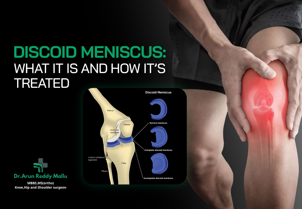
Discoid Meniscus: What It Is and How It’s Treated The knee is one of the most complex and important joints in the human body, allowing us to walk, run, squat, and jump. Inside the knee are two crescent-shaped pieces of cartilage known as the menisci — the medial (inner) and lateral (outer) meniscus. These structures act as shock absorbers between the thigh bone (femur) and the shin bone (tibia), distributing weight and providing stability. However, in some individuals, particularly children and adolescents, the meniscus may develop in an abnormal, disc-like shape rather than the typical crescent form. This condition is called a Discoid Meniscus. Though it can exist without symptoms, it often leads to pain, swelling, and mechanical issues in the knee, especially during activity. Let’s explore what a discoid meniscus is, its symptoms, causes, diagnosis, and treatment options available — including advanced care approaches offered by Dr. Arun Reddy Mallu at Continental Hospital, Hyderabad. What Is a Discoid Meniscus? A Discoid Meniscus is a congenital (present at birth) variation in which the meniscus is abnormally shaped like a disc rather than a crescent. This thicker, wider cartilage can cover a larger portion of the tibial surface than normal. The condition most commonly affects the lateral meniscus (outer part of the knee) and is more prevalent in children and young adults. In some cases, the discoid meniscus is more prone to tearing or becoming unstable because its shape and attachment to the surrounding tissues are abnormal. This makes it less flexible and more vulnerable during twisting or high-impact activities. Types of Discoid Meniscus Discoid menisci are classified into three main types based on their shape and attachment pattern: Incomplete Discoid Meniscus:The meniscus is partially disc-shaped and covers more of the tibial surface than usual but not entirely. Complete Discoid Meniscus:The meniscus completely covers the tibial surface and is thicker and more prone to tears. Wrisberg Variant (Hypermobile Meniscus):This type lacks normal posterior attachments, leading to excessive movement of the meniscus. It often causes “snapping” sensations in the knee. Causes of Discoid Meniscus The exact cause of a discoid meniscus isn’t fully understood, but it is believed to result from an abnormal development of the meniscus during fetal growth. Normally, the meniscus begins as a disc and reshapes into a crescent during early development. In cases of discoid meniscus, this reshaping process doesn’t occur completely. Although it is congenital, the condition might not present symptoms until later in childhood or adolescence when physical activity increases or a tear occurs. There is also a genetic predisposition, as discoid meniscus can sometimes run in families. Symptoms of Discoid Meniscus Many people with a discoid meniscus may not experience symptoms, and the condition is often discovered incidentally during an MRI or surgery for another knee issue. However, when symptoms do appear, they may include: Knee pain(especially during movement or twisting) Swellingor stiffness around the joint Popping or clicking sounds Locking or catching sensation Instabilityor “giving way” of the knee Limited range of motion A torn discoid meniscus can cause more severe symptoms, such as sharp pain, swelling, and an inability to fully straighten or bend the knee. How Is Discoid Meniscus Diagnosed? Early diagnosis is essential to prevent long-term damage to the knee joint. Your doctor will begin by taking a detailed medical history and performing a physical examination. During the exam, they may check for clicking, tenderness, range of motion, and joint stability. Imaging Tests: X-rays:While an X-ray doesn’t show the meniscus directly, it helps rule out bone injuries or abnormalities. MRI (Magnetic Resonance Imaging):The most effective diagnostic tool for identifying a discoid meniscus. MRI provides detailed images of the cartilage, showing its shape, thickness, and any associated tears or inflammation. Arthroscopy:In some cases, an arthroscopic procedure may be used both for diagnosis and treatment. It involves inserting a small camera into the knee jointfor a direct view of the meniscus. Treatment Options for Discoid Meniscus The treatment approach depends on several factors — such as the type of discoid meniscus, severity of symptoms, age, and level of activity. Non-Surgical Management If the discoid meniscus is asymptomatic or only mildly symptomatic, non-surgical treatments may be recommended, including: Rest and Activity Modification:Avoiding activities that cause pain or clicking. Physical Therapy:Strengthening the muscles around the knee improves stability and joint function. Medications:Over-the-counter pain relievers or anti-inflammatory drugs can help reduce swelling and discomfort. Regular Monitoring:Periodic follow-ups with imaging to ensure the condition is not worsening. Non-surgical management works best when the discoid meniscus is stable and there are no tears. Surgical Treatment If symptoms persist or if there is a meniscal tear, surgery may be required. Modern surgical techniques aim to preserve as much of the meniscus as possible, as complete removal can lead to early arthritis. The main surgical procedures include: Arthroscopic Saucerization (Meniscoplasty) This is the most common and preferred surgical method. The surgeon reshapes the discoid meniscus into a normal, crescent form, preserving its function while reducing the risk of future tears. Meniscal Repair If the meniscus has torn, the surgeon may repair it using sutures. Preserving the tissue is especially important in younger patients to maintain joint cushioning. Partial Meniscectomy If the tear is severe or in a non-repairable zone, the damaged portion may be trimmed away while leaving the rest intact. Total Meniscectomy In rare and extreme cases, when the meniscus is too damaged to save, the entire meniscus may be removed. However, this is avoided whenever possible because it increases the risk of arthritis over time. Recovery After Surgery Recovery depends on the type of procedure performed, but most patients can expect the following stages: Immediate Post-Operative Care:Ice, elevation, and prescribed pain medications help reduce swelling. Rehabilitation and Physical Therapy:Exercises begin soon after surgery to restore strength, flexibility, and range of motion. Return to Activities:Light activities may resume within a few weeks, while sports or heavy exercises typically resume after 3–6 months under medical guidance. Your orthopedic surgeon will tailor a rehabilitation plan based on your specific needs and surgery type. Potential Complications While most